Explore
 Validate
 Learn
Learn Western blot
 Immunocytochemistry
Antibody data
- Antibody Data
- Antigen structure
- References [4]
- Comments [0]
- Validations
- Western blot [7]
- Immunocytochemistry [1]
- Immunoprecipitation [1]
- Immunohistochemistry [1]
- Chromatin Immunoprecipitation [2]
Submit
Validation data
Reference
Comment
Report error
- Product number
- GTX112864 - Provider product page

- Provider
- GeneTex
- Proper citation
- GeneTex Cat#GTX112864, RRID:AB_11173565
- Product name
- PARP antibody [N2C1], Internal
- Antibody type
- Polyclonal
- Reactivity
- Human, Mouse, Rat
- Host
- Rabbit
Submitted references A high-throughput pipeline for validation of antibodies.
GSK3β negatively regulates TRAX, a scaffold protein implicated in mental disorders, for NHEJ-mediated DNA repair in neurons.
Reactive oxygen species-driven mitochondrial injury induces apoptosis by teroxirone in human non-small cell lung cancer cells.
The CHAC1-inhibited Notch3 pathway is involved in temozolomide-induced glioma cytotoxicity.
Sikorski K, Mehta A, Inngjerdingen M, Thakor F, Kling S, Kalina T, Nyman TA, Stensland ME, Zhou W, de Souza GA, Holden L, Stuchly J, Templin M, Lund-Johansen F
Nature methods 2018 Nov;15(11):909-912
Nature methods 2018 Nov;15(11):909-912
GSK3β negatively regulates TRAX, a scaffold protein implicated in mental disorders, for NHEJ-mediated DNA repair in neurons.
Chien T, Weng YT, Chang SY, Lai HL, Chiu FL, Kuo HC, Chuang DM, Chern Y
Molecular psychiatry 2018 Dec;23(12):2375-2390
Molecular psychiatry 2018 Dec;23(12):2375-2390
Reactive oxygen species-driven mitochondrial injury induces apoptosis by teroxirone in human non-small cell lung cancer cells.
Wang JP, Hsieh CH, Liu CY, Lin KH, Wu PT, Chen KM, Fang K
Oncology letters 2017 Sep;14(3):3503-3509
Oncology letters 2017 Sep;14(3):3503-3509
The CHAC1-inhibited Notch3 pathway is involved in temozolomide-induced glioma cytotoxicity.
Chen PH, Shen WL, Shih CM, Ho KH, Cheng CH, Lin CW, Lee CC, Liu AJ, Chen KC
Neuropharmacology 2017 Apr;116:300-314
Neuropharmacology 2017 Apr;116:300-314
No comments: Submit comment
Enhanced validation
Supportive validation
- Submitted by
- GeneTex (provider)
- Enhanced method
- Genetic validation
- Main image
- Experimental details
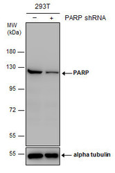
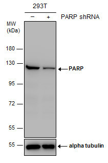
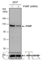
- Non-transfected (¡V) and transfected (+) 293T whole cell extracts (30 ?g) were separated by 7.5% SDS-PAGE, and the membrane was blotted with PARP antibody [N2C1], Internal (GTX112864) diluted at 1:5000. The HRP-conjugated anti-rabbit IgG antibody (GTX213110-01) was used to detect the primary antibody.
Supportive validation
- Submitted by
- GeneTex (provider)
- Main image
- Experimental details
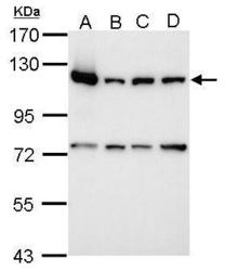
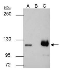
- Sample (30 ?g of whole cell lysate) A: 293T B: A431 C: HeLa D: HepG2 7.5% SDS PAGE GTX112864 diluted at 1:10000 The HRP-conjugated anti-rabbit IgG antibody (GTX213110-01) was used to detect the primary antibody.

- Submitted by
- GeneTex (provider)
- Main image
- Experimental details
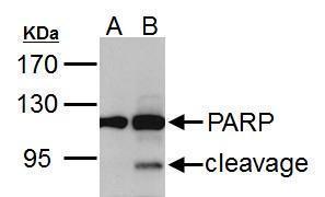
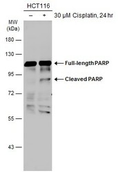
- PARP1 antibody [N2C1], Internal detects PARP1 protein by western blot analysis.A. 30 ?g HCT116 whole cell lysate/extract (untreated) B. 30 ?g HCT116 whole cell lysate/extract (30 ?M cisplatin treatment for 24hr)7.5% SDS-PAGEPARP1 antibody [N2C1], Internal (GTX112864) dilution: 1:5000 The HRP-conjugated anti-rabbit IgG antibody (GTX213110-01) was used to detect the primary antibody.
- Submitted by
- GeneTex (provider)
- Main image
- Experimental details
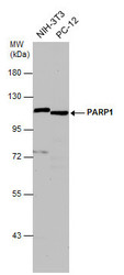
- Various whole cell extracts (30 ?g) were separated by 7.5% SDS-PAGE, and the membrane was blotted with PARP1 antibody [N2C1], Internal (GTX112864) diluted at 1:500. The HRP-conjugated anti-rabbit IgG antibody (GTX213110-01) was used to detect the primary antibody.
- Submitted by
- GeneTex (provider)
- Main image
- Experimental details
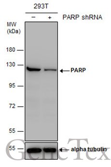
- Non-transfected (¡V) and transfected (+) 293T whole cell extracts (30 ?g) were separated by 7.5% SDS-PAGE, and the membrane was blotted with PARP antibody [N2C1], Internal (GTX112864) diluted at 1:5000. The HRP-conjugated anti-rabbit IgG antibody (GTX213110-01) was used to detect the primary antibody.
- Submitted by
- GeneTex (provider)
- Main image
- Experimental details
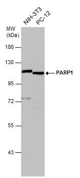
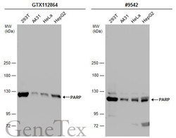
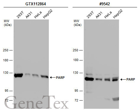
- Various whole cell extracts (30 ?g) were separated by 5% SDS-PAGE, and the membranes were blotted with PARP antibody [N2C1], Internal (GTX112864) diluted at 1:10000 and competitor's antibody (#9542) diluted at 1:500. The HRP-conjugated anti-rabbit IgG antibody (GTX213110-01) was used to detect the primary antibody.*The competitor is not affiliated with GeneTex and does not endorse this product.
- Submitted by
- GeneTex (provider)
- Main image
- Experimental details
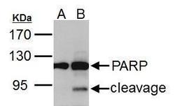
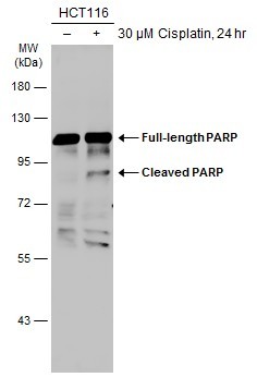
- Untreated (¡V) and treated (+) HCT-116 whole cell extract (30 ?g) were separated by 7.5% SDS-PAGE, and the membrane was blotted with PARP antibody (GTX112864) diluted at 1:1000. The HRP-conjugated anti-rabbit IgG antibody (GTX213110-01) was used to detect the primary antibody.
Supportive validation
- Submitted by
- GeneTex (provider)
- Main image
- Experimental details
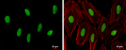
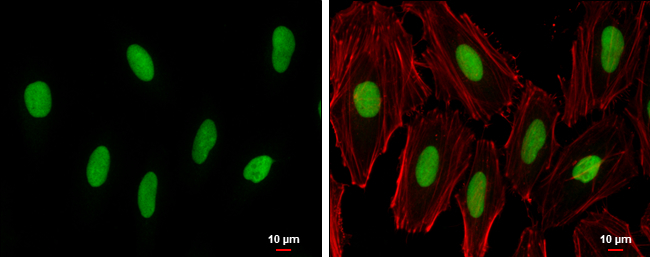
- PARP antibody [N2C1], Internal detects PARP protein at nucleus by immunofluorescent analysis.Sample: HeLa cells were fixed in 4% paraformaldehyde at RT for 15 min.Green: PARP protein stained by PARP antibody [N2C1], Internal (GTX112864) diluted at 1:500.Red: phalloidin, a cytoskeleton marker, diluted at 1:200.Blue: Hoechst 33342 staining.Scale bar = 10 £gm.
Supportive validation
- Submitted by
- GeneTex (provider)
- Main image
- Experimental details
- PARP1 antibody [N2C1], Internal immunoprecipitates PARP1 protein in IP experiments.IP samples: HCT-116 whole cell extractA. 30 £gg HCT-116 whole cell extractB. Control with 4 £gg of preimmune Rabbit IgGC. Immunoprecipitation of PARP1 protein by 4 £gg PARP1 antibody [N2C1], Internal (GTX112864)5 % SDS-PAGEThe immunoprecipitated PARP1 protein was detected by PARP1 antibody [N2C1], Internal (GTX112864) diluted at 1:500.[EasyBlot anti-rabbit IgG (GTX221666-01) was used as a secondary reagent]

Supportive validation
- Submitted by
- GeneTex (provider)
- Main image

- Experimental details
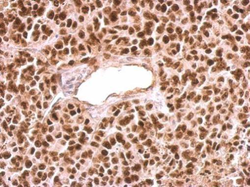
- PARP1 antibody [N2C1], Internal detects PARP1 protein at nucleus on HeLa xenograft by immunohistochemical analysis. Sample: Paraffin-embedded HeLa xenograft. PARP1 antibody [N2C1], Internal (GTX112864) dilution: 1:500.
Supportive validation
- Submitted by
- GeneTex (provider)
- Main image
- Experimental details
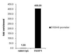
- Cross-linked ChIP was performed with Raji chromatin extract and 5 £gg of either control rabbit IgG or anti-PARP1 antibody. The precipitated DNA was detected by PCR with primer set targeting to S100A9 promoter.

- Submitted by
- GeneTex (provider)
- Main image
- Experimental details
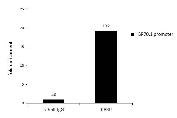
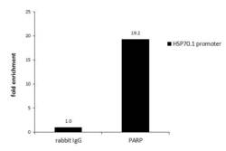
- ChIP was performed with HeLa chromatin extract and 5 ?g of either normal rabbit IgG or anti-PARP antibody. The precipitated DNA was detected by PCR with primer set targeting to HSP70.1 promoter.