 Explore
 Validate
 Learn
Learn Western blot
 Immunocytochemistry
 Immunohistochemistry
Antibody data
- Antibody Data
- Antigen structure
- References [3]
- Comments [0]
- Validations
- Western blot [1]
- Immunocytochemistry [1]
Submit
Validation data
Reference
Comment
Report error
- Product number
- AMAb90959 - Provider product page

- Provider
- Atlas Antibodies
- Proper citation
- Atlas Antibodies Cat#AMAb90959, RRID:AB_2665732
- Product name
- Anti-PARP1
- Antibody type
- Monoclonal
- Description
- Monoclonal Antibody against Human PARP1, Clone ID: CL2220, Gene description: poly (ADP-ribose) polymerase 1, Alternative Gene Names: ADPRT, PARP, PPOL, Validated applications: WB, IHC, ICC, Uniprot ID: P09874, Storage: Store at +4°C for short term storage. Long time storage is recommended at -20°C.
- Reactivity
- Human
- Host
- Mouse
- Conjugate
- Unconjugated
- Isotype
- IgG
- Antibody clone number
- CL2220
- Vial size
- 100 µl
- Concentration
- 1.0 mg/ml
- Storage
- Store at +4°C for short term storage. Long time storage is recommended at -20°C.
- Handling
- The antibody solution should be gently mixed before use.
Submitted references PARP targeted Auger emitter therapy with [125I]PARPi-01 for triple-negative breast cancer
Revealing protein-protein interactions at the transcriptome scale by sequencing.
RAD51-Mediated DNA Homologous Recombination Is Independent of PTEN Mutational Status
Ambur Sankaranarayanan R, Florea A, Allekotte S, Vogg A, Maurer J, Schäfer L, Bolm C, Terhorst S, Classen A, Bauwens M, Morgenroth A, Mottaghy F
EJNMMI Research 2022;12(1)
EJNMMI Research 2022;12(1)
Revealing protein-protein interactions at the transcriptome scale by sequencing.
Johnson KL, Qi Z, Yan Z, Wen X, Nguyen TC, Zaleta-Rivera K, Chen CJ, Fan X, Sriram K, Wan X, Chen ZB, Zhong S
Molecular cell 2021 Oct 7;81(19):4091-4103.e9
Molecular cell 2021 Oct 7;81(19):4091-4103.e9
RAD51-Mediated DNA Homologous Recombination Is Independent of PTEN Mutational Status
Sinha A, Saleh A, Endersby R, Yuan S, Chokshi C, Brown K, Kuzio B, Kauppinen T, Singh S, Baker S, McKinnon P, Katyal S
Cancers 2020;12(11):3178
Cancers 2020;12(11):3178
No comments: Submit comment
Enhanced validation
- Submitted by
- Atlas Antibodies (provider)
- Enhanced method
- Genetic validation
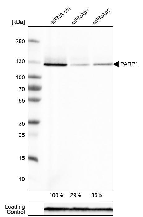
- Main image

- Experimental details
- Western blot analysis in RT-4 cells transfected with control siRNA, target specific siRNA probe #1 and #2, using Anti-PARP1 antibody. Remaining relative intensity is presented. Loading control: Anti-GAPDH.
- Sample type
- Human
- Protocol
- Protocol
Supportive validation
- Submitted by
- Atlas Antibodies (provider)
- Main image
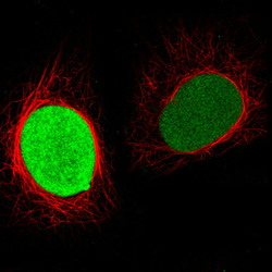
- Experimental details
- Immunofluorescence staining in HeLa cell line with Anti-PARP1 monoclonal antibody, showing cell cycle dependent nuclear staining in green. Microtubule- and nuclear probes are visualized in red and blue respectively (where available).
- Sample type
- Human
