Explore
 Validate
 Learn
Learn Western blot
 Immunocytochemistry
Antibody data
- Antibody Data
- Antigen structure
- References [1]
- Comments [0]
- Validations
- Immunocytochemistry [3]
- Immunoprecipitation [1]
- Immunohistochemistry [1]
- Chromatin Immunoprecipitation [4]
- Other assay [2]
Submit
Validation data
Reference
Comment
Report error
- Product number
- PA5-34802 - Provider product page

- Provider
- Invitrogen Antibodies
- Product name
- PARP1 Polyclonal Antibody
- Antibody type
- Polyclonal
- Antigen
- Recombinant full-length protein
- Description
- Recommended positive controls: 293T, A431, HeLa, HepG2, A549, PC-12, Rat2. Predicted reactivity: Mouse (84%), Rat (85%). Store product as a concentrated solution. Centrifuge briefly prior to opening the vial.
- Reactivity
- Human, Rat
- Host
- Rabbit
- Isotype
- IgG
- Vial size
- 100 μL
- Concentration
- 1 mg/mL
- Storage
- Store at 4°C short term. For long term storage, store at -20°C, avoiding freeze/thaw cycles.
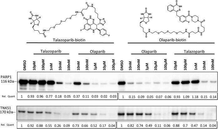
Submitted references Dissecting the molecular determinants of clinical PARP1 inhibitor selectivity for tankyrase1.
Ryan K, Bolaňos B, Smith M, Palde PB, Cuenca PD, VanArsdale TL, Niessen S, Zhang L, Behenna D, Ornelas MA, Tran KT, Kaiser S, Lum L, Stewart A, Gajiwala KS
The Journal of biological chemistry 2021 Jan-Jun;296:100251
The Journal of biological chemistry 2021 Jan-Jun;296:100251
No comments: Submit comment
Supportive validation
- Submitted by
- Invitrogen Antibodies (provider)
- Main image
- Experimental details
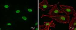
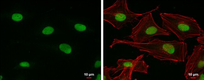
- Immunocytochemistry-Immunofluorescence analysis of PARP1 was performed in HeLa cells fixed in 4% paraformaldehyde at RT for 15 min. Green: PARP1 Polyclonal Antibody (Product # PA5-34802) diluted at 1:500. Red: phalloidin, a cytoskeleton marker. Scale bar = 10 µm.
- Submitted by
- Invitrogen Antibodies (provider)
- Main image
- Experimental details
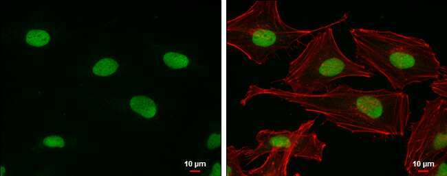
- Immunocytochemistry-Immunofluorescence analysis of PARP1 was performed in HeLa cells fixed in 4% paraformaldehyde at RT for 15 min. Green: PARP1 Polyclonal Antibody (Product # PA5-34802) diluted at 1:500. Red: phalloidin, a cytoskeleton marker. Scale bar = 10 µm.

- Submitted by
- Invitrogen Antibodies (provider)
- Main image

- Experimental details
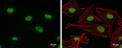
- Immunocytochemistry-Immunofluorescence analysis of PARP1 was performed in HeLa cells fixed in 4% paraformaldehyde at RT for 15 min. Green: PARP1 Polyclonal Antibody (Product # PA5-34802) diluted at 1:500. Red: phalloidin, a cytoskeleton marker. Scale bar = 10 µm.
Supportive validation
- Submitted by
- Invitrogen Antibodies (provider)
- Main image

- Experimental details
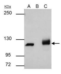
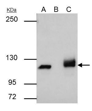
- PARP1 antibody [N1N2], N-term immunoprecipitates PARP1 protein in IP experiments. IP samples: HCT-116 whole cell extract. A. 30 µg HCT-116 whole cell extract. B. Control with 4 µg of preimmune Rabbit IgG. C. Immunoprecipitation of PARP1 protein by 4 µg PARP1 antibody [N1N2], N-term (Product # PA5-34802). 5 % SDS-PAGE. The immunoprecipitated PARP1 protein was detected by PARP1 antibody [N1N2], N-term (Product # PA5-34802) diluted at 1:500.

Supportive validation
- Submitted by
- Invitrogen Antibodies (provider)
- Main image
- Experimental details
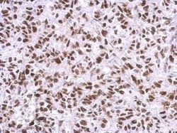
- Immunohistochemical analysis of paraffin-embedded HeLa xenograft, using PARP1 antibody [N1N2], N-term (Product # PA5-34802) antibody at 1:500 dilution. Antigen Retrieval: EDTA based buffer, pH 8.0, 15 min.

Supportive validation
- Submitted by
- Invitrogen Antibodies (provider)
- Main image

- Experimental details
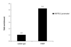
- ChIP assay analysis of PARP1 was performed in HeLa chromatin extract using 5 µg of either normal rabbit IgG or PARP1 Polyclonal Antibody (Product # PA5-34802). The precipitated DNA was detected by PCR with primer set targeting to HSP70.1 promoter.

- Submitted by
- Invitrogen Antibodies (provider)
- Main image
- Experimental details
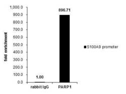
- Cross-linked ChIP was performed with Raji chromatin extract and 5 µg of either control rabbit IgG or PARP1 Polyclonal Antibody (Product # PA5-34802). The precipitated DNA was detected by PCR with primer set targeting to S100A9 promoter.
- Submitted by
- Invitrogen Antibodies (provider)
- Main image

- Experimental details
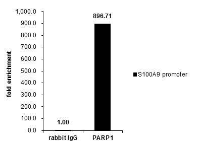
- Cross-linked ChIP was performed with Raji chromatin extract and 5 µg of either control rabbit IgG or PARP1 Polyclonal Antibody (Product # PA5-34802). The precipitated DNA was detected by PCR with primer set targeting to S100A9 promoter.

- Submitted by
- Invitrogen Antibodies (provider)
- Main image
- Experimental details
- ChIP assay analysis of PARP1 was performed in HeLa chromatin extract using 5 µg of either normal rabbit IgG or PARP1 Polyclonal Antibody (Product # PA5-34802). The precipitated DNA was detected by PCR with primer set targeting to HSP70.1 promoter.

Supportive validation
- Submitted by
- Invitrogen Antibodies (provider)
- Main image
- Experimental details
- PARP1 antibody [N1N2], N-term immunoprecipitates PARP1 protein in IP experiments. IP samples: HCT-116 whole cell extract. A. 30 µg HCT-116 whole cell extract. B. Control with 4 µg of preimmune Rabbit IgG. C. Immunoprecipitation of PARP1 protein by 4 µg PARP1 antibody [N1N2], N-term (Product # PA5-34802). 5 % SDS-PAGE. The immunoprecipitated PARP1 protein was detected by PARP1 antibody [N1N2], N-term (Product # PA5-34802) diluted at 1:500.
- Submitted by
- Invitrogen Antibodies (provider)
- Main image
- Experimental details
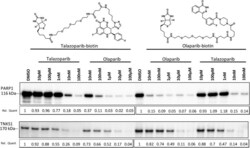
- Figure 1 Talazoparib binds to TNKS1 at a clinically relevant concentration in cultured cells. NCI-H1048 cell lysate was pretreated with indicated concentrations of talazoparib or olaparib (30 min) prior to the addition of talazoparib- or olaparib-biotin probes (30 min). Proteins bound to streptavidin were released and subjected to Western blot analysis for either PARP1 ( top ) or TNKS1 ( bottom ). Relative quantitation (Rel Quant) was determined using ImageJ and is displayed below the plots. PARP1, poly(ADP-ribosyl) polymerase 1; TNKS1, tankyrase1.