Explore
 Validate
 Learn
Learn Western blot
 Immunocytochemistry
Antibody data
- Antibody Data
- Antigen structure
- References [1]
- Comments [0]
- Validations
- Western blot [3]
- Immunocytochemistry [1]
- Immunoprecipitation [1]
Submit
Validation data
Reference
Comment
Report error
- Product number
- GTX632388 - Provider product page

- Provider
- GeneTex
- Product name
- PARP antibody [GT982]
- Antibody type
- Monoclonal
- Reactivity
- Human
- Host
- Mouse
Submitted references A high-throughput pipeline for validation of antibodies.
Sikorski K, Mehta A, Inngjerdingen M, Thakor F, Kling S, Kalina T, Nyman TA, Stensland ME, Zhou W, de Souza GA, Holden L, Stuchly J, Templin M, Lund-Johansen F
Nature methods 2018 Nov;15(11):909-912
Nature methods 2018 Nov;15(11):909-912
No comments: Submit comment
Enhanced validation
Supportive validation
- Submitted by
- GeneTex (provider)
- Enhanced method
- Genetic validation
- Main image
- Experimental details
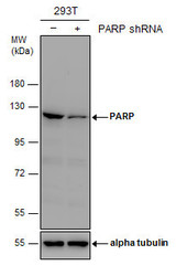
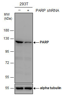
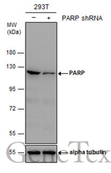
- Non-transfected (¡V) and transfected (+) 293T whole cell extracts (30 ?g) were separated by 7.5% SDS-PAGE, and the membrane was blotted with PARP antibody [GT982] (GTX632388) diluted at 1:1000.
Supportive validation
- Submitted by
- GeneTex (provider)
- Main image
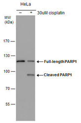
- Experimental details
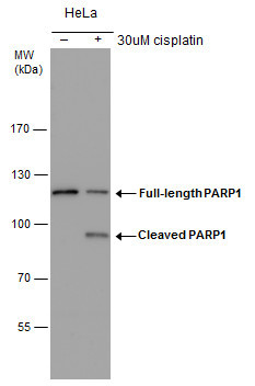
- PARP1 antibody detects PARP1 protein by western blot analysis. Un-treated (-) and treated (+, 30uM cisplatin treatment for 24hr) HeLa whole cell extracts (30 £gg) were separated by 7.5% SDS-PAGE, and the membrane was blotted with PARP1 antibody (GTX632388) at a dilution of 1:1000.
- Submitted by
- GeneTex (provider)
- Main image
- Experimental details
- Non-transfected (¡V) and transfected (+) 293T whole cell extracts (30 ?g) were separated by 7.5% SDS-PAGE, and the membrane was blotted with PARP antibody [GT982] (GTX632388) diluted at 1:1000.

Supportive validation
- Submitted by
- GeneTex (provider)
- Main image
- Experimental details
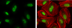
- PARP1 antibody [GT982] detects PARP1 protein at nucleus by immunofluorescent analysis.Sample: HeLa cells were fixed in 4% paraformaldehyde at RT for 15 min.Green: PARP1 protein stained by PARP1 antibody [GT982] (GTX632388) diluted at 1:400.Red: Phalloidin, a F-actin marker, diluted at 1:200.

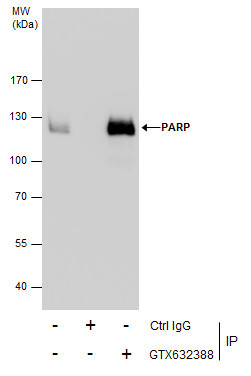
Supportive validation
- Submitted by
- GeneTex (provider)
- Main image
- Experimental details
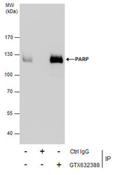
- Immunoprecipitation of PARP protein from 293T whole cell extracts using 5 £gg of PARP antibody (GTX632388).Western blot analysis was performed using PARP antibody (GTX632388).EasyBlot anti-Mouse IgG (GTX221667-01) was used as a secondary reagent.