 Explore
 Validate
 Learn
Learn Western blot
Antibody data
- Antibody Data
- Antigen structure
- References [0]
- Comments [0]
- Validations
- Western blot [2]
- Immunocytochemistry [1]
- Flow cytometry [1]
Submit
Validation data
Reference
Comment
Report error
- Product number
- 702343 - Provider product page

- Provider
- Invitrogen Antibodies
- Product name
- PARP1 Recombinant Rabbit Monoclonal Antibody (8H2L9)
- Antibody type
- Monoclonal
- Antigen
- Synthetic peptide
- Reactivity
- Human
- Host
- Rabbit
- Isotype
- IgG
- Antibody clone number
- 8H2L9
- Vial size
- 100 µg
- Concentration
- 0.5 mg/mL
- Storage
- Store at 4°C short term. For long term storage, store at -20°C, avoiding freeze/thaw cycles.
No comments: Submit comment
Supportive validation
- Submitted by
- Invitrogen Antibodies (provider)
- Main image
- Experimental details
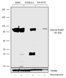
- Western blot analysis was performed on nuclear cell extracts (30 µg lysate) of Jurkat (Lane 1), Jurkat treated with Staurosporine (1uM overnight) (Lane 2), NTERA-2 (Lane 3), NTERA-2 treated with Staurosporine (1uM overnight) (Lane 4), SH-SY5Y (Lane 5) and SH-SY5Y treated with Staurosporine (1uM overnight) (Lane 6). The blots were probed with Anti-Cleaved PARP Recombinant Rabbit Monoclonal Antibody (Product # 702343, 1-2 µg/mL) and detected by chemiluminescence using Goat anti-Rabbit IgG (H+L) Superclonal™ Secondary Antibody, HRP conjugate (Product # A27036, 0.4 µg/mL, 1:2500 dilution). A 89 kDa band corresponding to Cleaved PARP was observed and the minor fragment at 35 kDa was observed only in NTERA-2 cells treated with Staurosporine. The antibody does not detect the full length PARP but is specific to the cleaved form. Known quantity of protein samples were electrophoresed using Novex® NuPAGE® 4-12% Bis-Tris gel (Product # NP0321BOX), XCell SureLock™ Electrophoresis System (Product # EI0002) and Novex® Sharp Pre-Stained Protein Standard (Product # LC5800). Resolved proteins were then transferred onto a nitrocellulose membrane with iBlot® Dry Blotting System (Product # IB21001). The membrane was probed with the relevant primary and secondary Antibody following blocking with 5% skimmed milk. Chemiluminescent detection was performed using Pierce™ ECL Western blotting Substrate (Product # 32106).

- Submitted by
- Invitrogen Antibodies (provider)
- Main image
- Experimental details
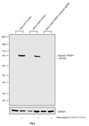
- CRISPR-Cas9 mediated genome editing ofPARP1 (as confirmed by next generation sequencing) was achieved by using LentiArray™ Lentiviral sgRNA (Product # A32042, AssayID CRISPR978664_LV) and LentiArray Cas9 Lentivirus (Product # A32064). Fig (a) Western blot analysis of PARP1 was performed by loading 30 µg of HeLa Wild Type (Lane 1), HeLa Wild Type treated with Staurosporin (1 µM for 16hrs) (Lane 2), HeLa Cas9 (Lane 3), HeLa Cas9 treated with Staurosporin (1 µM for 16hrs) (Lane 4), HeLa Cas9 cells transduced with PARP1 Lentiviral sgRNA (Lane 5) and HeLa Cas9 cells transduced with PARP1 Lentiviral sgRNA treated with Staurosporin (1 µM for 16hrs) (Lane 6) whole cell extracts. The samples were electrophoresed using NuPAGE™ Novex™ 4-12% Bis-Tris Protein Gel (Product # NP0322BOX). Resolved proteins were then transferred onto a nitrocellulose membrane (Product # IB23001) by iBlot® 2 Dry Blotting System (Product # IB21001). The blot was probed with Anti-PARP1 Recombinant Rabbit Monoclonal Antibody (8H2L9) (Product # 702343) using 1:500 dilution and Goat anti-Rabbit IgG (H+L) Superclonal™ Recombinant Secondary Antibody, HRP (Product # A27036 1:2,500 dilution).Chemiluminescent detection was performed using Novex® ECL Chemiluminescent Substrate Reagent Kit (Product # WP20005). Loss of signal in sgRNA transduced cells using the LentiArray™ CRISPR product line confirms that antibody is specific to PARP1.

Supportive validation
- Submitted by
- Invitrogen Antibodies (provider)
- Main image

- Experimental details
- For immunofluorescence analysis, Staurosporine (1 uM, 16 h) treated HeLa cells were fixed and permeabilized for detection of endogenous PARP cleavage using Anti-Cleaved PARP beginning at N214 Recombinant Rabbit Monoclonal Antibody (Product # 702343, 2 µg/mL) and labeled with Goat anti-Rabbit IgG (H+L) Superclonal™ Secondary Antibody, Alexa Fluor® 488 conjugate (Product # A27034, 1:2000). Panel a) shows representative cells that were stained for detection and localization of Cleaved PARP protein (green), Panel b) is stained for nuclei (blue) using SlowFade® Gold Antifade Mountant with DAPI (Product # S36938). Panel c) represents cytoskeletal F-actin staining using Alexa Fluor® 555 Rhodamine Phalloidin (Product # R415, 1:300). Panel d) is a composite image of Panels a, b and c clearly demonstrating nuclear localization of Cleaved PARP. Panel e) shows untreated cells with no signal. Panel f) represents control cells with no primary antibody to assess background. The images were captured at 60X magnification.

Supportive validation
- Submitted by
- Invitrogen Antibodies (provider)
- Main image

- Experimental details
- Flow Cytometry analysis of endogenous Cleaved PARP was performed on Jurkat cells (untreated, red histogram) and Jurkat cells treated with 1 uM staurosporine for 16 h (blue histogram), labeled with ABfinity™ Anti-Cleaved PARP Recombinant Rabbit Monoclonal Antibody (Product # 702343, 1 uM/ 1M cells) or with rabbit isotype control at 0.5 ug/ml (pink histogram) and detected with Goat anti-Rabbit IgG (H+L) Superclonal™ Secondary Antibody, (Alexa Fluor® 488 conjugate, Product # A27034, 0.4 ug/ml, 1:2500). The purple histogram represents unstained control cells. A representative of 10,000 cells were acquired and analyzed for each sample using an Attune® Acoustic Focusing Cytometer (4468770).
