 Explore
 Validate
 Learn
Learn Western blot
Antibody data
- Antibody Data
- Antigen structure
- References [6]
- Comments [0]
- Validations
- Western blot [1]
- Immunocytochemistry [1]
Submit
Validation data
Reference
Comment
Report error
- Product number
- 14-6667-82 - Provider product page

- Provider
- Invitrogen Antibodies
- Product name
- PARP1 Monoclonal Antibody (HC2R8), eBioscience™
- Antibody type
- Monoclonal
- Antigen
- Other
- Description
- Description: This HC2R8 monoclonal antibody reacts with human poly (ADP-ribose) polymerase (PARP1). This ubiquitous 116 kDa nuclear enzyme is involved in DNA repair. During apoptosis, active caspases -3, -6 and -7 cleave PARP1 after Asp214, thereby inactivating PARP1 and generating two apoptotic fragments sized 85 kDa and 25 kDa. The HC2R8 antibody specifically recognizes both the cleaved and non-cleaved forms of PARP. Applications Reported: This HC2R8 antibody has been reported for use in immunoblotting (WB). Applications Tested: This HC2R8 antibody has been tested by immunoblotting of staurosporine-treated Jurkat cells. This can be used at less than or equal to 0.1 µg/mL. It is recommended that the antibody be carefully titrated for optimal performance in the assay of interest. Purity: Greater than 90%, as determined by SDS-PAGE. Aggregation: Less than 10%, as determined by HPLC. Filtration: 0.2 µm post-manufacturing filtered.
- Reactivity
- Human
- Host
- Mouse
- Isotype
- IgG
- Antibody clone number
- HC2R8
- Vial size
- 100 µg
- Concentration
- 0.5 mg/mL
- Storage
- 4° C
Submitted references Identification of an acetylation-dependant Ku70/FLIP complex that regulates FLIP expression and HDAC inhibitor-induced apoptosis.
The role of proteases during apoptosis.
Yama/CPP32 beta, a mammalian homolog of CED-3, is a CrmA-inhibitable protease that cleaves the death substrate poly(ADP-ribose) polymerase.
Structural and functional analysis of poly(ADP ribose) polymerase: an immunological study.
Production and characterization of monoclonal antibodies specific for the functional domains of poly(ADP-ribose) polymerase.
Poly(ADP-ribose) polymerase is a zinc metalloenzyme.
Kerr E, Holohan C, McLaughlin KM, Majkut J, Dolan S, Redmond K, Riley J, McLaughlin K, Stasik I, Crudden M, Van Schaeybroeck S, Fenning C, O'Connor R, Kiely P, Sgobba M, Haigh D, Johnston PG, Longley DB
Cell death and differentiation 2012 Aug;19(8):1317-27
Cell death and differentiation 2012 Aug;19(8):1317-27
The role of proteases during apoptosis.
Patel T, Gores GJ, Kaufmann SH
FASEB journal : official publication of the Federation of American Societies for Experimental Biology 1996 Apr;10(5):587-97
FASEB journal : official publication of the Federation of American Societies for Experimental Biology 1996 Apr;10(5):587-97
Yama/CPP32 beta, a mammalian homolog of CED-3, is a CrmA-inhibitable protease that cleaves the death substrate poly(ADP-ribose) polymerase.
Tewari M, Quan LT, O'Rourke K, Desnoyers S, Zeng Z, Beidler DR, Poirier GG, Salvesen GS, Dixit VM
Cell 1995 Jun 2;81(5):801-9
Cell 1995 Jun 2;81(5):801-9
Structural and functional analysis of poly(ADP ribose) polymerase: an immunological study.
Lamarre D, Talbot B, de Murcia G, Laplante C, Leduc Y, Mazen A, Poirier GG
Biochimica et biophysica acta 1988 Jul 13;950(2):147-60
Biochimica et biophysica acta 1988 Jul 13;950(2):147-60
Production and characterization of monoclonal antibodies specific for the functional domains of poly(ADP-ribose) polymerase.
Lamarre D, Talbot B, Leduc Y, Muller S, Poirier G
Biochemistry and cell biology = Biochimie et biologie cellulaire 1986 Apr;64(4):368-76
Biochemistry and cell biology = Biochimie et biologie cellulaire 1986 Apr;64(4):368-76
Poly(ADP-ribose) polymerase is a zinc metalloenzyme.
Zahradka P, Ebisuzaki K
European journal of biochemistry 1984 Aug 1;142(3):503-9
European journal of biochemistry 1984 Aug 1;142(3):503-9
No comments: Submit comment
Supportive validation
- Submitted by
- Invitrogen Antibodies (provider)
- Main image

- Experimental details
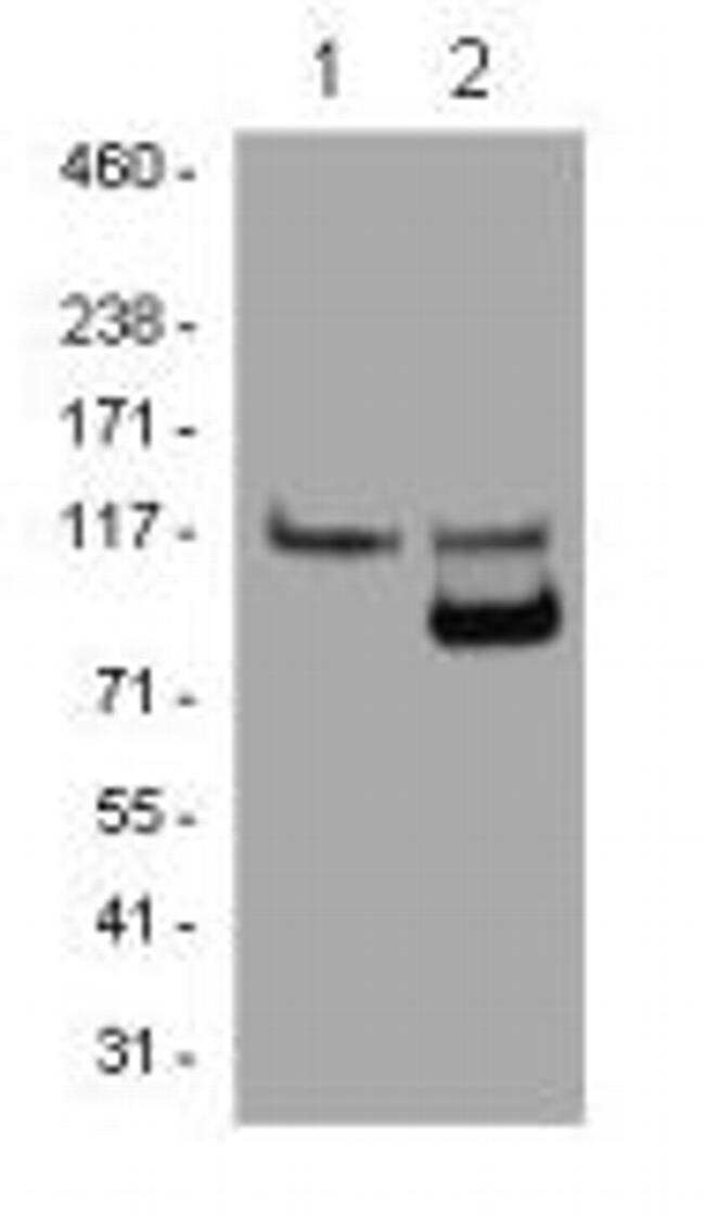
- Cell lysates prepared from Jurkat cells left untreated (lane 1) or treated for 2 hrs with staurosporine (lane 2) were immunoblotted with 0.1 µg/mL of the Anti-Human PARP1 antibody. Bands were visualized using Anti-Mouse IgG HRP.
Supportive validation
- Submitted by
- Invitrogen Antibodies (provider)
- Main image
- Experimental details
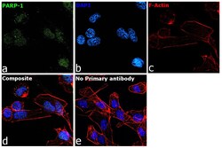
- Immunofluorescence analysis of PARP1 was performed using 70% confluent log phase MDA-MB-231 cells. The cells were fixed with 4% paraformaldehyde for 10 minutes, permeabilized with 0.1% Triton™ X-100 for 15 minutes, and blocked with 2% BSA for 45 minutes at room temperature. The cells were labeled with PARP1 Monoclonal Antibody (HC2R8), eBioscience™ (Product # 14-6667-82) at 5µg/mL in 0.1% BSA, incubated at 4 degree celsius overnight and then labeled with Goat anti-Mouse IgG (H+L) Highly Cross-Adsorbed Secondary Antibody, Alexa Fluor Plus 488 (Product # A32723, 1:2000), for 45 minutes at room temperature (Panel a: Green). Nuclei (Panel b:Blue) were stained with ProLong™ Diamond Antifade Mountant with DAPI (Product # P36962). F-actin (Panel c: Red) was stained with Rhodamine Phalloidin (Product # R415, 1:300). Panel d represents the merged image showing nuclear localization. Panel e represents control cells with no primary antibody to assess background. The images were captured at 60X magnification.
