Explore
 Validate
 Learn
Learn Western blot
Antibody data
- Antibody Data
- Antigen structure
- References [0]
- Comments [0]
- Validations
- Western blot [5]
- Immunohistochemistry [1]
Submit
Validation data
Reference
Comment
Report error
- Product number
- LS-C745005 - Provider product page

- Provider
- LSBio
- Product name
- PARP1 Antibody (N-Terminus) LS-C745005
- Antibody type
- Polyclonal
- Description
- Affinity chromatography
- Reactivity
- Human
- Host
- Rabbit
- Isotype
- IgG
- Storage
- Store vial at -20°C or below prior to opening. Dilute 1:10 to minimize loss. Store the vial at -20°C or below after dilution. Avoid freeze-thaw cycles.
No comments: Submit comment
Supportive validation
- Submitted by
- LSBio (provider)
- Enhanced method
- Genetic validation
- Main image

- Experimental details
- Western Blot of rabbit anti-PARP1 N-term Antibody. Lane 1: Opal Pre-stained ladder Lane 2: OVCAR-8 Wild Type. Lane 3: PARP1-KO. Lane 4: PARP2-KO. Lane 5: PARP3-KO. Lane 6: PARP4-KO Lane 7: PARP5a-KO. Lane 8: PARP5b-KO. Lane 9: PARP6-KO. Lane 10: PARP7-KO. Lane 11: PARP8-KO. Lane 12: PARP9-KO. Lane 13: PARP10-KO. Lane 14: PARP12-KO. Lane 15: PARP13-KO. Lane 16: PARP14-KO. Lane 17: PARP16-KO. Load: 5.0 µg per lane. Primary antibody: PARP1 n-term antibody at 1ug/mL overnight at 4°C. Secondary antibody: Goat anti-rabbit Peroxidase secondary antibody at 1:40,000 for 30 min at RT. Blocking Buffer: MB-073 for 30 min at RT. Predicted/Observed size: ~113 kDa for PARP1.

- Submitted by
- LSBio (provider)
- Enhanced method
- Genetic validation
- Main image

- Experimental details
- Western Blot of rabbit anti-PARP1 antibody. Marker: Opal Pre-stained ladder Lane 1: HEK293 lysate Lane 2: HeLa Lysate Lane 3: MCF-7 Lysate Lane 4: Jurkat Lysate Lane 5: A431 Lysate Lane 6: A549 Lysate Lane 7: LNCap Lysate Lane 8: MOLT-4 Lysate Lane 9: Ramos Lysate Lane 10: Raji Lsyate Lane 11: A-172 Lysate Lane 12: NIH/3T3 Lysate Load: 35 µg per lane. Primary antibody: PARP1 antibody at 1ug/mL overnight at 4C. Secondary antibody: Peroxidase rabbit secondary antibody at 1:30,000 for 60 min at RT. Blocking Buffer: 1% Casein-TTBS for 30 min at RT. Predicted/Observed size: 113 kDa for PARP1.

- Submitted by
- LSBio (provider)
- Enhanced method
- Genetic validation
- Main image

- Experimental details
- Western Blot of endogenous PARP1 with Rabbit Anti-PARP1 (N-term ZF1) Antibody. Lane 1: OVCAR8 Wild Type lysate. Lane 2: OVCAR8 PARP1 KO lysate. Load: 5 µg per lane. Primary antibody: PARP1 (N-term ZF1) antibody at 1µg/mL for overnight at 4°C. Secondary antibody: HRP Gt-a-Rb IgG secondary antibody at 1:40,000 for 30 min at RT. Block: MB-070 overnight at 4°C. Predicted/Observed size: 113 kDa for endogenous PARP1. Other band(s): none. Image in collaboration with Phil Lorenzi at MD Anderson.

- Submitted by
- LSBio (provider)
- Enhanced method
- Genetic validation
- Main image

- Experimental details
- Western Blot of recombinant PARP1 with rabbit anti-PARP1 (N-term ZF1) antibody. Lane 1: PARP1-Zinc Finger domain recombinant protein. Load: 0.05 µg per lane. Primary antibody: PARP1 (N-term ZF1) antibody at 1µg/mL for overnight at 4°C. Secondary antibody: HRP Gt-a-rabbit secondary antibody at 1:40,000 for 30 min at RT. Block: MB-070 overnight at 4°C. Predicted/Observed size: 13 kDa for rPARP1 (N-term ZF1). Other band(s): none.

- Submitted by
- LSBio (provider)
- Enhanced method
- Genetic validation
- Main image

- Experimental details
- Western Blot of endogenous PARP1 with Rabbit Anti-PARP1 Antibodies. Lane 1: OVCAR8 Wild Type lysate. Lane 2: OVCAR8 PARP1 KO lysate. Load 5 ? per lane. Primary Antibody: Blot A: Anti-PARP1- n term; Blot B: Anti-PARP1- internal at 1?/mL for overnight at 4?. Secondary antibody: HRP Gt-a-Rb IgG secondary antibody at 1:40,000 for 30 min at RT. Block: MB-070 overnight at 4?. Predicted/Observed size: 113 kDa for endogenous PARP1. Other band(s): nonspecific ~ 40kDa in PARP1-AD only.

Supportive validation
- Submitted by
- LSBio (provider)
- Main image

- Experimental details
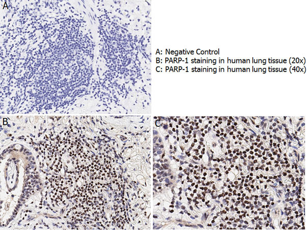
- Immunohistochemistry with anti-PARP-1 antibody showing nuclear positivity in human lung tissue at 20x and 40x (B & C). Staining was performed on Leica Bond system using the standard protocol. Formalin fixed/paraffin embedded tissue sections were subjected to antigen retrieval and then incubated with rabbit anti-PARP-1 antibody at 1:100 dilution for 60 minutes. Biotinylated Anti-rabbit secondary antibody was used at 1:200 dilution to detect primary antibody. The reaction was developed using streptavidin-HRP conjugated compact polymer system and visualized with chromogen substrate, 3?-diamino-benzidine substrate (DAB). The sections were then counterstained with hematoxylin to detect cell nuclei.