Explore
 Validate
 Learn
Learn Western blot
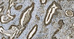
 Immunocytochemistry
Antibody data
- Antibody Data
- Antigen structure
- References [2]
- Comments [0]
- Validations
- Western blot [1]
Submit
Validation data
Reference
Comment
Report error
- Product number
- M00122-5 - Provider product page

- Provider
- Boster Biological Technology
- Product name
- Anti-PARP Picoband™ Antibody (monoclonal, 10E11)
- Antibody type
- Monoclonal
- Description
- Mouse IgG monoclonal antibody for PARP detection. Tested with WB, IHC-P, ICC/IF, FCM in Human;Mouse;Rat.
- Reactivity
- Human, Mouse, Rat
- Host
- Mouse
- Isotype
- IgG
- Antibody clone number
- 1.10E+02
- Vial size
- 100μg/vial
- Concentration
- Add 0.2ml of distilled water will yield a concentration of 500ug/ml.
- Storage
- At -20°C for one year. After reconstitution, at 4°C for one month. It can also be aliquotted and stored frozen at -20°C for a longer time. Avoid repeated freezing and thawing.
- Handling
- Add 0.2ml of distilled water will yield a concentration of 500ug/ml.
Submitted references Inotodiol inhibits cells migration and invasion and induces apoptosis via p53-dependent pathway in HeLa cells.
Treatment with MQA, a Derivative of Caffeoylquinic Acid, Provides Neuroprotective Effects against Cerebral Ischemia Through Suppression of the p38 Pathway and Oxidative Stress in Rats.
Zhang SD, Yu L, Wang P, Kou P, Li J, Wang LT, Wang W, Yao LP, Zhao XH, Fu YJ
Phytomedicine : international journal of phytotherapy and phytopharmacology 2019 Jul;60:152957
Phytomedicine : international journal of phytotherapy and phytopharmacology 2019 Jul;60:152957
Treatment with MQA, a Derivative of Caffeoylquinic Acid, Provides Neuroprotective Effects against Cerebral Ischemia Through Suppression of the p38 Pathway and Oxidative Stress in Rats.
Chen L, Liu DN, Wang Y, Liu XY, Han S, Zhang K, Li GY, Tian X, Wang HY, Wang JH
Journal of molecular neuroscience : MN 2019 Apr;67(4):604-612
Journal of molecular neuroscience : MN 2019 Apr;67(4):604-612
No comments: Submit comment
Supportive validation
- Submitted by
- Boster Biological Technology (provider)
- Main image
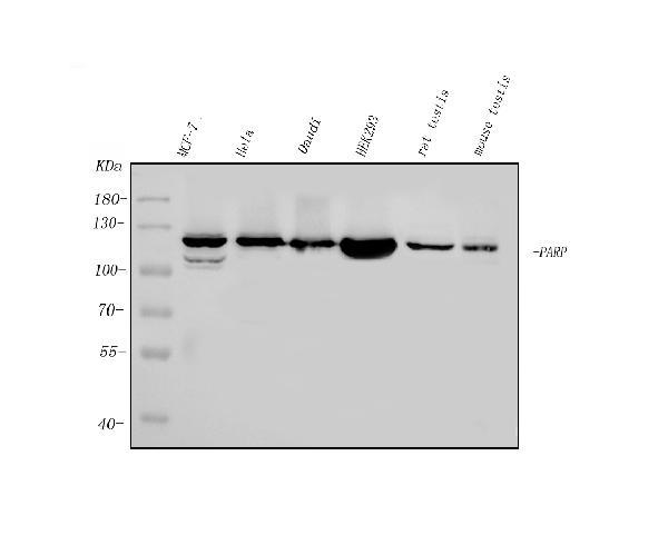
- Experimental details
- Western blot analysis of PARP using anti-PARP antibody (M00122-5). Electrophoresis was performed on a 5-20% SDS-PAGE gel at 70V (Stacking gel) / 90V (Resolving gel) for 2-3 hours. The sample well of each lane was loaded with 50ug of sample under reducing conditions. Lane 1: human MCF-7 whole cell lysates, Lane 2: human HELA whole cell lysates, Lane 3: human DAUDI whole cell lysates, Lane 4: human HEK293 whole cell lysates, Lane 5: rat testis tissue lysates, Lane 6: mouse testis tissue lysates. After Electrophoresis, proteins were transferred to a Nitrocellulose membrane at 150mA for 50-90 minutes. Blocked the membrane with 5% Non-fat Milk/ TBS for 1.5 hour at RT. The membrane was incubated with mouse anti- PARP antigen affinity purified monoclonal antibody (Catalog # M00122-5) at 0.25 μg/mL overnight at 4°C, then washed with TBS-0.1%Tween 3 times with 5 minutes each and probed with a goat anti-mouse IgG-HRP secondary antibody at a dilution of 1:10000 for 1.5 hour at RT. The signal is developed using an Enhanced Chemiluminescent detection (ECL) kit (Catalog # EK1001) with Tanon 5200 system. A specific band was detected for PARP at approximately 113KD. The expected band size for PARP is at 113KD.
- Additional image