Explore
 Validate
 Learn
Learn Western blot
 Immunohistochemistry
Antibody data
- Antibody Data
- Antigen structure
- References [8]
- Comments [0]
- Validations
- Western blot [2]
- Immunocytochemistry [2]
Submit
Validation data
Reference
Comment
Report error
- Product number
- HPA045168 - Provider product page

- Provider
- Atlas Antibodies
- Proper citation
- Atlas Antibodies Cat#HPA045168, RRID:AB_2679240
- Product name
- Anti-PARP1
- Antibody type
- Polyclonal
- Description
- Polyclonal Antibody against Human PARP1, Gene description: poly (ADP-ribose) polymerase 1, Alternative Gene Names: ADPRT, PARP, PPOL, Validated applications: WB, IHC, ICC, Uniprot ID: P09874, Storage: Store at +4°C for short term storage. Long time storage is recommended at -20°C.
- Reactivity
- Human
- Host
- Rabbit
- Conjugate
- Unconjugated
- Isotype
- IgG
- Vial size
- 100 µl
- Concentration
- 0.2 mg/ml
- Storage
- Store at +4°C for short term storage. Long time storage is recommended at -20°C.
- Handling
- The antibody solution should be gently mixed before use.
Submitted references Depletion of the RNA-binding protein PURA triggers changes in posttranscriptional gene regulation and loss of P-bodies
Human TRMT2A methylates tRNA and contributes to translation fidelity
Poly ADP-ribosylation of SET8 leads to aberrant H4K20 methylation in mammalian nuclear genome
Correlation between molar activity, injection mass and uptake of the PARP targeting radiotracer [(18)F]olaparib in mouse models of glioma.
NSG-Pro mouse model for uncovering resistance mechanisms and unique vulnerabilities in human luminal breast cancers
[18F]AZD2461, an Insight on Difference in PARP Binding Profiles for DNA Damage Response PET Imaging
YWHAZ amplification/overexpression defines aggressive bladder cancer and contributes to chemo‐/radio‐resistance by suppressing caspase‐mediated apoptosis
Poly (ADP-Ribose) Polymerase 1 Mediated Arginase II Activation Is Responsible for Oxidized LDL-Induced Endothelial Dysfunction
Molitor L, Klostermann M, Bacher S, Merl-Pham J, Spranger N, Burczyk S, Ketteler C, Rusha E, Tews D, Pertek A, Proske M, Busch A, Reschke S, Feederle R, Hauck S, Blum H, Drukker M, Fischer-Posovszky P, König J, Zarnack K, Niessing D
Nucleic Acids Research 2023;51(3):1297-1316
Nucleic Acids Research 2023;51(3):1297-1316
Human TRMT2A methylates tRNA and contributes to translation fidelity
Witzenberger M, Burczyk S, Settele D, Mayer W, Welp L, Heiss M, Wagner M, Monecke T, Janowski R, Carell T, Urlaub H, Hauck S, Voigt A, Niessing D
Nucleic Acids Research 2023;51(16):8691-8710
Nucleic Acids Research 2023;51(16):8691-8710
Poly ADP-ribosylation of SET8 leads to aberrant H4K20 methylation in mammalian nuclear genome
Estève P, Sen S, Vishnu U, Ruse C, Chin H, Pradhan S
Communications Biology 2022;5(1)
Communications Biology 2022;5(1)
Correlation between molar activity, injection mass and uptake of the PARP targeting radiotracer [(18)F]olaparib in mouse models of glioma.
Chan CY, Hopkins SL, Guibbal F, Pacelli A, Baguña Torres J, Mosley M, Lau D, Isenegger P, Chen Z, Wilson TC, Dias G, Hueting R, Gouverneur V, Cornelissen B
EJNMMI research 2022 Oct 9;12(1):67
EJNMMI research 2022 Oct 9;12(1):67
NSG-Pro mouse model for uncovering resistance mechanisms and unique vulnerabilities in human luminal breast cancers
Sun Y, Yang N, Utama F, Udhane S, Zhang J, Peck A, Yanac A, Duffey K, Langenheim J, Udhane V, Xia G, Peterson J, Jorns J, Nevalainen M, Rouet R, Schofield P, Christ D, Ormandy C, Rosenberg A, Chervoneva I, Tsaih S, Flister M, Fuchs S, Wagner K, Rui H
Science Advances 2021;7(38)
Science Advances 2021;7(38)
[18F]AZD2461, an Insight on Difference in PARP Binding Profiles for DNA Damage Response PET Imaging
Guibbal F, Hopkins S, Pacelli A, Isenegger P, Mosley M, Torres J, Dias G, Mahaut D, Hueting R, Gouverneur V, Cornelissen B
Molecular Imaging and Biology 2020;22(5):1226-1234
Molecular Imaging and Biology 2020;22(5):1226-1234
YWHAZ amplification/overexpression defines aggressive bladder cancer and contributes to chemo‐/radio‐resistance by suppressing caspase‐mediated apoptosis
Yu C, Li C, Chen I, Lai M, Lin Z, Korla P, Chai C, Ko G, Chen C, Hwang T, Lee S, Sheu J
The Journal of Pathology 2019;248(4):476-487
The Journal of Pathology 2019;248(4):476-487
Poly (ADP-Ribose) Polymerase 1 Mediated Arginase II Activation Is Responsible for Oxidized LDL-Induced Endothelial Dysfunction
Wang Q, Zhao T, Zhang W, Yu W, Liu B, Wang Z, Qiao W, Lu Q, Wang A, Zhang M
Frontiers in Pharmacology 2018;9
Frontiers in Pharmacology 2018;9
No comments: Submit comment
Enhanced validation
Enhanced validation
- Submitted by
- klas2
- Enhanced method
- Genetic validation
- Main image
- Experimental details
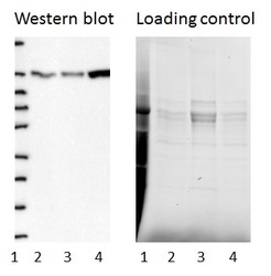
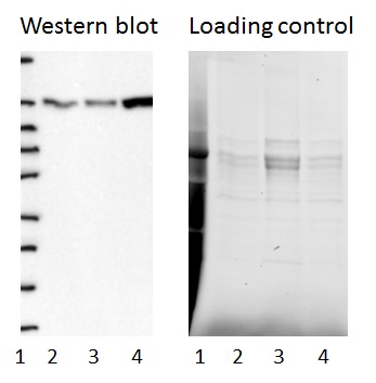
- Western blot of cell lysate from U-2 OS cells transfected with either siRNA targeting PARP1 or control siRNA. Lane 1: Marker (250, 130, 95, 72, 55, 36, 28, 17, 10) Lane 2: Cell lysate from U-2OS cells transfected with siRNA targeting PARP1 Lane 3: N/A Lane 4: Cell lysate from U-2OS cells transfected with control siRNA Right image, lane 1-4: loading control
- Sample type
- U-2 OS
- Primary Ab dilution
- 1:180
- Conjugate
- Horseradish Peroxidase
- Secondary Ab
- Secondary Ab
- Secondary Ab dilution
- 1:3000
- Knockdown/Genetic Approaches Application
- Western blot
Enhanced validation
- Submitted by
- Atlas Antibodies (provider)
- Enhanced method
- Genetic validation
- Main image

- Experimental details
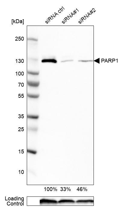
- Western blot analysis in RT-4 cells transfected with control siRNA, target specific siRNA probe #1 and #2, using Anti-PARP1 antibody. Remaining relative intensity is presented. Loading control: Anti-GAPDH.
- Sample type
- Human
- Protocol
- Protocol
Enhanced validation
Supportive validation
- Submitted by
- 55af80e3e0991
- Enhanced method
- Genetic validation
- Main image
- Experimental details
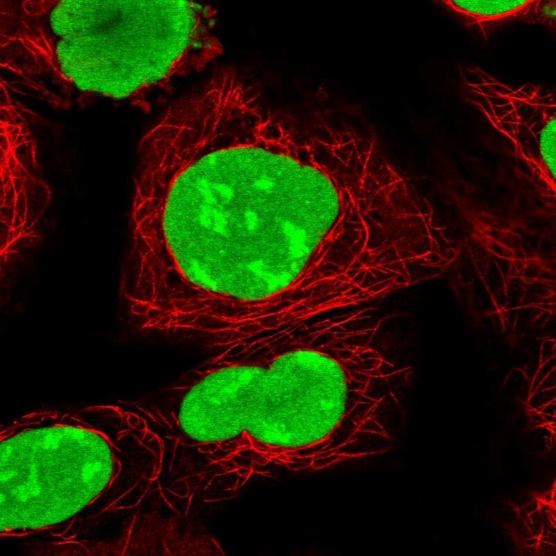
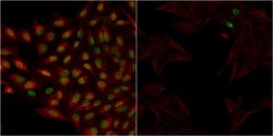
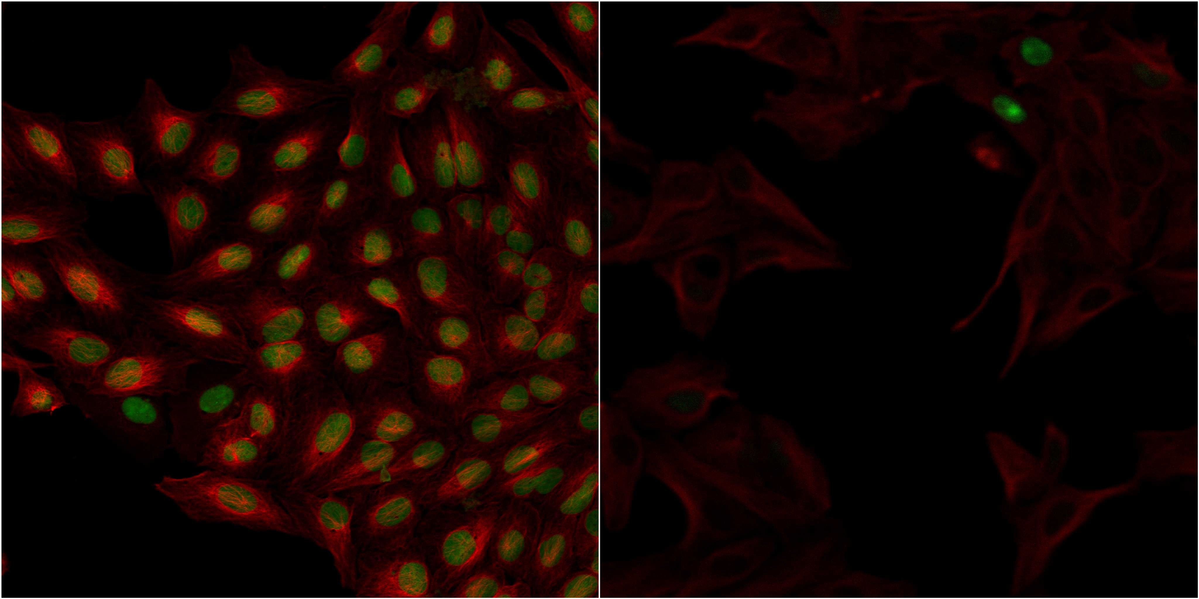
- Confocal images of immunofluorescently stained human U-2 OS cells.The protein PARP1 is shown in green and the microtubules in red. The image to the left show cells transfected with control siRNA and the image to the right show cells where PARP1 has been downregulated with specific siRNA.
- Sample type
- U-2 OS cells
- Primary Ab dilution
- 1:81
- Secondary Ab
- Secondary Ab
- Secondary Ab dilution
- 1:800
- Knockdown/Genetic Approaches Application
- Immunocytochemistry
Supportive validation
- Submitted by
- Atlas Antibodies (provider)
- Main image
- Experimental details
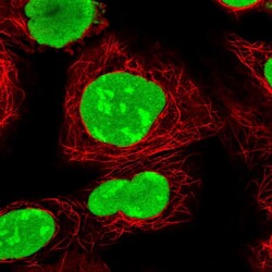
- Immunofluorescent staining of human cell line HEK 293 shows localization to nucleus.
- Sample type
- Human