 Explore
 Validate
 Learn
Learn Western blot
 Flow cytometry
Antibody data
- Antibody Data
- Antigen structure
- References [0]
- Comments [0]
- Validations
- Western blot [4]
- Immunocytochemistry [1]
Submit
Validation data
Reference
Comment
Report error
- Product number
- MA1-41020 - Provider product page

- Provider
- Invitrogen Antibodies
- Product name
- PARP1 (cleaved Asp214, Asp215) Monoclonal Antibody (194C1439)
- Antibody type
- Monoclonal
- Antigen
- Synthetic peptide
- Description
- Suggested positive control: camptothecin treated HL-60 (12 hr).
- Reactivity
- Human, Mouse
- Host
- Mouse
- Isotype
- IgG
- Antibody clone number
- 194C1439
- Vial size
- 100 µg
- Concentration
- 1.0 mg/mL
- Storage
- Store at 4°C short term. For long term storage, store at -20°C, avoiding freeze/thaw cycles.
No comments: Submit comment
Supportive validation
- Submitted by
- Invitrogen Antibodies (provider)
- Main image

- Experimental details
- Western blot analysis of cleaved PARP in staurosporine-treated Jurkat cells at various time points, using a PARP monoclonal antibody (Product # MA1-41020) at 2 µg/mL. The band corresponding to cleaved PARP is only seen in the treated samples.

- Submitted by
- Invitrogen Antibodies (provider)
- Main image

- Experimental details
- Western blot analysis of PARP1 (cleaved Asp214, Asp215) in staurosporine-treated Jurkat cells. Samples were incubated in PARP1 (cleaved Asp214, Asp215) monoclonal antibody (Product # MA1-41020) using a dilution of 2 µg/mL followed by an Anti-mouse Ig HRP conjugate secondary antibody. The band corresponding to cleaved PARP is only seen in the treated samples.

- Submitted by
- Invitrogen Antibodies (provider)
- Main image

- Experimental details
- Western blot was performed using Anti-PARP Monoclonal Antibody (194C1439) (Product # MA1-41020) and a 85 kDa band corresponding to PARP (cleaved form) was observed across all cell lines along with an uncharacterized band (*) at ~50 kDa. Nuclear enriched extracts (40 µg lysate) of Jurkat (Lane 1), Jurkat treated with Etoposide (1 µM for 16 hours) (Lane 2), HeLa (Lane 3), HeLa treated with Etoposide (1 µM for 16 hours) (Lane 4), HeLa (Lane 5), HeLa treated with Staurosporine (3 µM for 16 hours) (Lane 6) were electrophoresed using NuPAGE™ 4-12% Bis-Tris Protein Gel (Product # NP0321BOX). Resolved proteins were then transferred onto a Nitrocellulose membrane (Product # IB23001) by iBlot® 2 Dry Blotting System (Product # IB21001). The blot was probed with the primary antibody (1 µg/mL concentration) and detected by chemiluminescence with Goat anti-Mouse IgG (H+L) Superclonal™ Recombinant Secondary Antibody, HRP (Product # A28177,1:4000 dilution) using the iBright FL 1000 (Product # A32752). Chemiluminescent detection was performed using SuperSignal™ West Dura Extended Duration Substrate (Product # 34076).

- Submitted by
- Invitrogen Antibodies (provider)
- Main image

- Experimental details
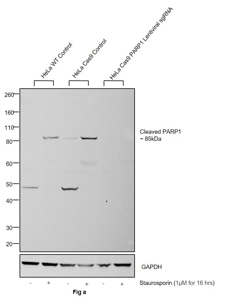
- CRISPR-Cas9 mediated genome editing of PARP1 (as confirmed by next generation sequencing) was achieved by using LentiArray™ Lentiviral sgRNA (Product # A32042, Assay ID CRISPR978664_LV) and LentiArray Cas9 Lentivirus (Product # A32064). Western blot analysis of PARP1 was performed by loading 30 µg of HeLa Wild Type (Lane 1), Treated HeLa Wild Type (Lane 2), HeLa Cas9 (Lane 3), Treated HeLa Cas9 (Lane 4), HeLa Cas9 cells transduced with PARP1 Lentiviral sgRNA (Lane 5) and Treated HeLa Cas9 cells transduced with PARP1 Lentiviral sgRNA (Lane 6) whole cell extracts. The samples were electrophoresed using NuPAGE™ Novex™ 4-12% Bis-Tris Protein Gel (Product # NP0322BOX). Resolved proteins were then transferred onto a nitrocellulose membrane (Product # IB23001) by iBlot® 2 Dry Blotting System (Product # IB21001). The blot was probed with Anti-PARP1 (cleaved Asp214, Asp215) Monoclonal Antibody (194C1439) (Product # MA1-41020) using 1:1,000 dilution and Goat anti-Mouse IgG (H+L) Superclonal™ Recombinant Secondary Antibody, HRP (Product # A28177 1:4,000 dilution). Chemiluminescent detection was performed using Novex® ECL Chemiluminescent Substrate Reagent Kit (Product # WP20005). Even though NGS analysis determine the clone as partial KO, there was complete loss of signal in sgRNA transduced cells confirming that the antibody is specific to PARP1. An uncharacterized band was observed at 49 kDa in HeLa Wild Type and Cas9 untreated samples. Treatment
Supportive validation
- Submitted by
- Invitrogen Antibodies (provider)
- Main image

- Experimental details
- Immunocytochemistry analysis of PARP1 (cleaved Asp214, Asp215) in A431 cells fixed for 10 minutes using 10% formalin and then permeabilized for 5 minutes using 1X PBS + 0.5% Triton-X100. Samples were incubated in PARP1 (cleaved Asp214, Asp215) monoclonal antibody (Product # MA1-41020) using a dilution of 5 µg/mL overnight at 4 °C followed by anti-mouse DyLight 488 (Green) with a dilution of 1:500. Nuclei were counterstained with DAPI (Blue). Cells were imaged using a 40X objective.
