 Explore
 Validate
 Learn
Learn Western blot
 Immunoprecipitation
Antibody data
- Antibody Data
- Antigen structure
- References [2]
- Comments [0]
- Validations
- Western blot [3]
- Immunocytochemistry [2]
- Chromatin Immunoprecipitation [1]
- Other assay [1]
Submit
Validation data
Reference
Comment
Report error
- Product number
- MA5-15031 - Provider product page

- Provider
- Invitrogen Antibodies
- Product name
- PARP1 Monoclonal Antibody (C.384.8)
- Antibody type
- Monoclonal
- Antigen
- Synthetic peptide
- Description
- It is not recommended to aliquot this antibody. This antibody is not cross-reactive with PARP-2 and PARP-3.
- Reactivity
- Human, Mouse, Rat
- Host
- Rabbit
- Isotype
- IgG
- Antibody clone number
- C.384.8
- Vial size
- 100 µL
- Concentration
- 438 µg/mL
- Storage
- -20°C
Submitted references A Novel Role for the DNA Repair Enzyme 8-Oxoguanine DNA Glycosylase in Adipogenesis.
Radiation induces EIF2AK3/PERK and ERN1/IRE1 mediated pro-survival autophagy.
Komakula SSB, Blaze B, Ye H, Dobrzyn A, Sampath H
International journal of molecular sciences 2021 Jan 25;22(3)
International journal of molecular sciences 2021 Jan 25;22(3)
Radiation induces EIF2AK3/PERK and ERN1/IRE1 mediated pro-survival autophagy.
Chaurasia M, Gupta S, Das A, Dwarakanath BS, Simonsen A, Sharma K
Autophagy 2019 Aug;15(8):1391-1406
Autophagy 2019 Aug;15(8):1391-1406
No comments: Submit comment
Supportive validation
- Submitted by
- Invitrogen Antibodies (provider)
- Main image

- Experimental details
- Western blot analysis of PARP in extracts from THP-1 cells, untreated or treated with TNF-alpha and cycloheximide using PARP monoclonal antibody (Product # MA5-15031).

- Submitted by
- Invitrogen Antibodies (provider)
- Main image

- Experimental details
- Western blot analysis was performed on modified whole cell extracts (1%SDS) (30 µg lysate) of HeLa (Lane 1), HeLa treated with Staurosporine (1uM for 12 hours) (Lane 2), Jurkat (Lane 3), Jurkat treated with Staurosporine (1 uM for 12 hours) (Lane 4), MCF7 (Lane 5), A549 (Lane 6), SH-SY5Y (Lane 7) and HT29 (Lane 8). The blot was probed with Anti-PARP Monoclonal Antibody (C.384.8) (Product # MA5-15031, 1:500 dilution) and detected by chemiluminescence using Goat anti-Rabbit IgG (H+L) Superclonal™ Secondary Antibody, HRP conjugate (Product # A27036, 0.25 µg/ml, 1:4000 dilution). A 113 kDa band corresponding to full length PARP and 86 kDa band corresponding to cleaved PARP was observed upon cell treatment in HeLa, Jurkat and was also observed in total cell lines like MCF7, A549, SH-SY5Y and HT29.

- Submitted by
- Invitrogen Antibodies (provider)
- Main image

- Experimental details
- CRISPR-Cas9 mediated genome editing ofPARP1 (as confirmed by next generation sequencing) was achieved by using LentiArray™ Lentiviral sgRNA (Product # A32042, Assay ID CRISPR978664_LV) and LentiArray Cas9 Lentivirus (Product # A32064). Western blot analysis of PARP1 was performed by loading 30 µg of HeLa Wild Type (Lane 1), HeLa Wild Type treated with Staurosporin (1 µM for 16 hrs) (Lane 2), HeLa Cas9 (Lane 3), HeLa Cas9 treated with Staurosporin (1 µM for 16 hrs) (Lane 4), HeLa Cas9 cells transduced with PARP1 Lentiviral sgRNA (Lane 5) and HeLa Cas9 cells transduced with PARP1 Lentiviral sgRNA treated with Staurosporin (1 µM for 16 hrs) (Lane 6) whole cell extracts. The samples were electrophoresed using NuPAGE™ Novex™ 4-12% Bis-Tris Protein Gel (Product # NP0322BOX). Resolved proteins were then transferred onto a nitrocellulose membrane (Product # IB23001) by iBlot® 2 Dry Blotting System (Product # IB21001). The blot was probed with Anti-PARP1 Monoclonal Antibody (C.384.8) (Product # MA5-15031) using 1:500 dilution and Goat anti-Rabbit IgG (H+L) Superclonal™ Recombinant Secondary Antibody, HRP (Product # A27036 1:4,000 dilution). Chemiluminescent detection was performed using Novex® ECL Chemiluminescent Substrate Reagent Kit (Product # WP20005). Even though NGS analysis determine the clone as partial KO, there was complete loss of signal in sgRNA transduced cells using the LentiArray™ CRISPR product line confirm

Supportive validation
- Submitted by
- Invitrogen Antibodies (provider)
- Main image

- Experimental details
- Immunofluorescent analysis of PARP (red) in HEK293T cells. Cells fixed with 4% formaldehyde were permeabilized and blocked with 1X PBS containing 5% BSA and 0.3% Triton X-100 for 1 hour at room temperature. Cells were probed with a PARP monoclonal antibody (Product # MA5-15031) at a dilution of 1:200 overnight at 4°C in 1X PBS containing 1% BSA and 0.3% Triton X-100, washed with 1X PBS, and incubated with a fluorophore-conjugated goat anti-rabbit IgG secondary antibody at a dilution of 1:200 for 1hr at room temperature. Nuclei (blue) were stained with DAPI. Images were taken on a Leica DM1000 microscope at 40X magnification. Data courtesy of the Innovators Program.

- Submitted by
- Invitrogen Antibodies (provider)
- Main image

- Experimental details
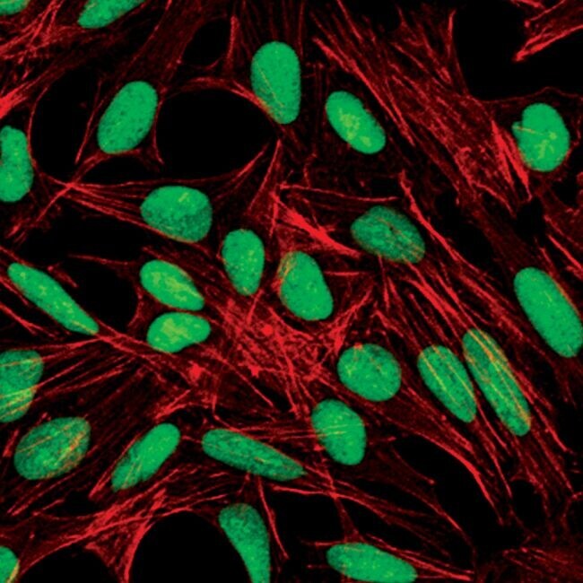
- Immunofluorescent analysis of PARP in untreated HeLa cells using a PARP monoclonal antibody (Product # MA5-15031) (green). Actin filaments are labeled with a fluorescent red phalloidin.
Supportive validation
- Submitted by
- Invitrogen Antibodies (provider)
- Main image

- Experimental details
- Enrichment of endogenous PARP protein at specific gene loci using Anti-PARP Antibody: Chromatin Immunoprecipitation (ChIP) was performed using Anti-PARP Rabbit Monoclonal Antibody (Product # MA5-15031, 8 µl) on sheared chromatin from 2 million HeLa cells treated with Etoposide (50 µM for 6 hours) using the MAGnify ChIP System (Product # 49-2024). Normal Rabbit IgG was used as a negative IP control. The purified DNA was analyzed by qPCR with PCR primer pairs over IGFBP6, IGFBP7, GAPDH promoter and gene, SAT2 satellite repeats and SAT alpha. Data is presented as fold enrichment of the antibody signal versus the negative control IgG using the comparative CT method.

Supportive validation
- Submitted by
- Invitrogen Antibodies (provider)
- Main image
- Experimental details
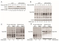
- Figure 5 OGG1 genotype alters PARylation of cellular proteins in adipocytes and adipose tissue. Proteins were isolated from 3T3-L1-CARDelta cells transduced with GFP or MTS-hOGG1-1a for detection of ( A ) PARP1 and ( B ) total cellular PARylation by immunoblotting. Cellular PARylation was assessed in protein extracts from differentiated primary adipocytes and adipose tissue from ( C ) WT vs. Ogg1 Tg animals and ( D ) from WT vs. Ogg1 -/- mice. Data are representative of at least 3 independent replicates for cell studies and 4-6 age- and sex-matched animals per genotype for tissue extracts. * p < 0.05 vs. 0 h.
