 Explore
 Validate
 Learn
Learn Western blot
Antibody data
- Antibody Data
- Antigen structure
- References [5]
- Comments [0]
- Validations
- Western blot [4]
- Immunocytochemistry [1]
- Immunohistochemistry [3]
- Other assay [1]
Submit
Validation data
Reference
Comment
Report error
- Product number
- PA5-16452 - Provider product page

- Provider
- Invitrogen Antibodies
- Product name
- PARP1 Polyclonal Antibody
- Antibody type
- Polyclonal
- Antigen
- Synthetic peptide
- Description
- PA5-16452 targets PARP (Poly ADP-Ribose Polymerase) in IHC (P) and WB applications and shows reactivity with mouse, Rat, and Human samples. The PA5-16452 immunogen is a synthetic peptide for the N-terminal region of human PARP.
- Reactivity
- Human, Mouse, Rat
- Host
- Rabbit
- Isotype
- IgG
- Vial size
- 500 µL
- Concentration
- 1 mg/mL
- Storage
- 4° C
Submitted references Improved radiosynthesis of (123)I-MAPi, an auger theranostic agent.
Validation of the use of a fluorescent PARP1 inhibitor for the detection of oral, oropharyngeal and oesophageal epithelial cancers.
Targeted Brain Tumor Radiotherapy Using an Auger Emitter.
Kaempferol Inhibits Zearalenone-Induced Oxidative Stress and Apoptosis via the PI3K/Akt-Mediated Nrf2 Signaling Pathway: In Vitro and In Vivo Studies.
Generation of Homogenous Three-Dimensional Pancreatic Cancer Cell Spheroids Using an Improved Hanging Drop Technique.
Wilson TC, Jannetti SA, Guru N, Pillarsetty N, Reiner T, Pirovano G
International journal of radiation biology 2023;99(1):70-76
International journal of radiation biology 2023;99(1):70-76
Validation of the use of a fluorescent PARP1 inhibitor for the detection of oral, oropharyngeal and oesophageal epithelial cancers.
Kossatz S, Pirovano G, Demétrio De Souza França P, Strome AL, Sunny SP, Zanoni DK, Mauguen A, Carney B, Brand C, Shah V, Ramanajinappa RD, Hedne N, Birur P, Sihag S, Ghossein RA, Gönen M, Strome M, Suresh A, Molena D, Ganly I, Kuriakose MA, Patel SG, Reiner T
Nature biomedical engineering 2020 Mar;4(3):272-285
Nature biomedical engineering 2020 Mar;4(3):272-285
Targeted Brain Tumor Radiotherapy Using an Auger Emitter.
Pirovano G, Jannetti SA, Carter LM, Sadique A, Kossatz S, Guru N, Demétrio De Souza França P, Maeda M, Zeglis BM, Lewis JS, Humm JL, Reiner T
Clinical cancer research : an official journal of the American Association for Cancer Research 2020 Jun 15;26(12):2871-2881
Clinical cancer research : an official journal of the American Association for Cancer Research 2020 Jun 15;26(12):2871-2881
Kaempferol Inhibits Zearalenone-Induced Oxidative Stress and Apoptosis via the PI3K/Akt-Mediated Nrf2 Signaling Pathway: In Vitro and In Vivo Studies.
Rajendran P, Ammar RB, Al-Saeedi FJ, Mohamed ME, ElNaggar MA, Al-Ramadan SY, Bekhet GM, Soliman AM
International journal of molecular sciences 2020 Dec 28;22(1)
International journal of molecular sciences 2020 Dec 28;22(1)
Generation of Homogenous Three-Dimensional Pancreatic Cancer Cell Spheroids Using an Improved Hanging Drop Technique.
Ware MJ, Colbert K, Keshishian V, Ho J, Corr SJ, Curley SA, Godin B
Tissue engineering. Part C, Methods 2016 Apr;22(4):312-21
Tissue engineering. Part C, Methods 2016 Apr;22(4):312-21
No comments: Submit comment
Supportive validation
- Submitted by
- Invitrogen Antibodies (provider)
- Main image
- Experimental details
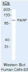
- Western blot of PARP (Poly ADP-Ribose Polymerase) using PARP (Poly ADP-Ribose Polymerase) Polyclonal Antibody (Product # PA5-16452) on Raji Cells.

- Submitted by
- Invitrogen Antibodies (provider)
- Main image
- Experimental details
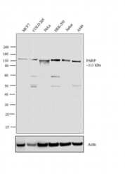
- Western blot analysis was performed on nuclear enriched extracts (30 µg lysate) of MCF7 (Lane 1), COLO 205 (Lane 2), HeLa (Lane 3), HEK-293 (Lane 4), Jurkat (Lane 5) and A549 (Lane 6). The blots were probed with Anti-PARP Rabbit Polyclonal Antibody (Product # PA5-16452, 2 µg/mL) and detected by chemiluminescence using Goat anti-Rabbit IgG (H+L) Superclonal™ Secondary Antibody, HRP conjµgate (Product # A27036, 0.4 µg/mL, 1:2500 dilution). A 113 kDa band corresponding to PARP was observed across the cell lines tested. Known quantity of protein samples were electrophoresed using Novex® NuPAGE® 10 % Bis-Tris gel (Product # NP0302BOX), XCell SureLock™ Electrophoresis System (Product # EI0002) and Novex® Sharp Pre-Stained Protein Standard (Product # LC5800). Resolved proteins were then transferred onto a nitrocellulose membrane with PierceTM Power Blotter System (Product # 22834). The membrane was probed with the relevant primary and secondary Antibody following blocking with 5 % skimmed milk. Chemiluminescent detection was performed using Pierce™ ECL Western Blotting Substrate (Product # 32106).

- Submitted by
- Invitrogen Antibodies (provider)
- Main image

- Experimental details
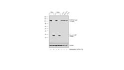
- CRISPR-Cas9 mediated genome editing ofPARP1 (as confirmed by next generation sequencing) was achieved by using LentiArray™ Lentiviral sgRNA (Product # A32042, AssayID CRISPR978664_LV) and LentiArray Cas9 Lentivirus (Product # A32064). Fig (a) Western blot analysis of PARP1 was performed by loading 30 µg of HeLa Cas9 (Lane 1), HeLa Cas9 treated with Staurosporin (1 µM for 16 hrs) (Lane 2), HeLa Cas9 cells transduced with PARP1 Lentiviral sgRNA (Lane 3) and HeLa Cas9 cells transduced with PARP1 Lentiviral sgRNA treated with Staurosporin (1 µM for 16 hrs) (Lane 4) whole cell extracts. The samples were electrophoresed using NuPAGE™ Novex™ 4-12% Bis-Tris Protein Gel (Product # NP0322BOX). Resolved proteins were then transferred onto a nitrocellulose membrane (Product # IB23001) by iBlot® 2 Dry Blotting System (Product # IB21001). The blot was probed with Anti-PARP1 Polyclonal Antibody (Product # PA5-16452) using 1:1,000 dilution and Goat anti-Rabbit IgG (H+L) Superclonal™ Recombinant Secondary Antibody, HRP (Product # A27036 1:4,000 dilution).Chemiluminescent detection was performed using Novex® ECL Chemiluminescent Substrate Reagent Kit (Product # WP20005). Loss of signal in sgRNA transduced cells using the LentiArray™ CRISPR product line confirms that antibody is specific to PARP1.

- Submitted by
- Invitrogen Antibodies (provider)
- Main image
- Experimental details
- Western blot analysis was performed on modified whole cell extracts (1%SDS) (30 µg lysate) of HeLa (Lane 1), HeLa treated with Staurosporine (1 µM for 12 hours) (Lane 2), Jurkat (Lane 3), Jurkat treated with Staurosporine (1 µM for 12 hours) (Lane 4), MCF7 (Lane 5), SH-SY5Y (Lane 6) and HT29 (Lane 7). The blot was probed with Anti-PARP Polyclonal Antibody (Product # PA5-16452, 1 µg/mL) and detected by chemiluminescence using Goat anti-Rabbit IgG (H+L) Superclonal™ Secondary Antibody, HRP conjugate (Product # A27036, 0.25 µg/mL, 1:4000 dilution). A 113 kDa band corresponding to full length PARP and 24 kDa band corresponding to cleaved PARP upon cell treatment was observed in HeLa, Jurkat and was also observed in total cell lines like MCF7, SH-SY5Y and HT29.

Supportive validation
- Submitted by
- Invitrogen Antibodies (provider)
- Main image

- Experimental details
- Immunofluorescence analysis of PARP was performed using 70% confluent log phase HeLa cells. The cells were fixed with 4% paraformaldehyde for 10 minutes, permeabilized with 0.1% Triton™ X-100 for 10 minutes, and blocked with 1% BSA for 1 hour at room temperature. The cells were labeled with PARP (Poly ADP-Ribose Polymerase) Rabbit Polyclonal Antibody (Product # PA5-16452) at 2µg/mL in 0.1% BSA and incubated for 3 hours at room temperature and then labeled with Goat anti-Rabbit IgG (H+L) Superclonal™ Secondary Antibody, Alexa Fluor® 488 conjµgate (Product # A27034) at a dilution of 1:2000 for 45 minutes at room temperature (Panel a: green). Nuclei (Panel b: blue) were stained with SlowFade® Gold Antifade Mountant with DAPI (Product # S36938). F-actin (Panel c: red) was stained with Alexa Fluor® 555 Rhodamine Phalloidin (Product # R415, 1:300). Panel d represents the merged image showing nuclear localization. Panel e shows the no primary antibody control. The images were captured at 60X magnification.

Supportive validation
- Submitted by
- Invitrogen Antibodies (provider)
- Main image

- Experimental details
- Formalin-fixed, paraffin-embedded human tonsil stained with PARP antibody using peroxidase-conjugate and AEC chromogen. Note nuclear staining of cells.

- Submitted by
- Invitrogen Antibodies (provider)
- Main image

- Experimental details
- Immunohistochemistry analysis of PARP (Poly ADP-Ribose Polymerase) showing staining in the nucleus of paraffin-embedded human tonsil tissue (right) compared to a negative control without primary antibody (left). To expose target proteins, antigen retrieval was performed using 10mM sodium citrate (pH 6.0), microwaved for 8-15 min. Following antigen retrieval, tissues were blocked in 3% H2O2-methanol for 15 min at room temperature, washed with ddH2O and PBS, and then probed with a PARP (Poly ADP-Ribose Polymerase) Rabbit Polyclonal Antibody (Product # PA5-16452) diluted in 3% BSA-PBS at a dilution of 1:20 for 1 hour at 37°C in a humidified chamber. Tissues were washed extensively in PBST and detection was performed using an HRP-conjugated secondary antibody followed by colorimetric detection using a DAB kit. Tissues were counterstained with hematoxylin and dehydrated with ethanol and xylene to prep for mounting.

- Submitted by
- Invitrogen Antibodies (provider)
- Main image

- Experimental details
- Immunohistochemistry analysis of PARP (Poly ADP-Ribose Polymerase) showing staining in the nucleus of paraffin-embedded mouse spleen tissue (right) compared to a negative control without primary antibody (left). To expose target proteins, antigen retrieval was performed using 10mM sodium citrate (pH 6.0), microwaved for 8-15 min. Following antigen retrieval, tissues were blocked in 3% H2O2-methanol for 15 min at room temperature, washed with ddH2O and PBS, and then probed with a PARP (Poly ADP-Ribose Polymerase) Rabbit Polyclonal Antibody (Product # PA5-16452) diluted in 3% BSA-PBS at a dilution of 1:20 for 1 hour at 37°C in a humidified chamber. Tissues were washed extensively in PBST and detection was performed using an HRP-conjugated secondary antibody followed by colorimetric detection using a DAB kit. Tissues were counterstained with hematoxylin and dehydrated with ethanol and xylene to prep for mounting.

Supportive validation
- Submitted by
- Invitrogen Antibodies (provider)
- Main image

- Experimental details
- Immunoprecipitation of PARP (Poly ADP-Ribose Polymerase) using PARP (Poly ADP-Ribose Polymerase) Polyclonal Antibody (Product # PA5-16452) on denatured Human Raji Cells.
