Explore
 Validate
 Learn
Learn Western blot
Antibody data
- Antibody Data
- Antigen structure
- References [3]
- Comments [0]
- Validations
- Western blot [3]
- Immunocytochemistry [1]
- Immunoprecipitation [1]
Submit
Validation data
Reference
Comment
Report error
- Product number
- AF-600-NA - Provider product page

- Provider
- R&D Systems
- Product name
- Human/Mouse PARP Antibody
- Antibody type
- Polyclonal
- Description
- Antigen Affinity-purified. Detects human and mouse PARP in Western blots.
- Reactivity
- Human, Mouse
- Host
- Goat
- Conjugate
- Unconjugated
- Antigen sequence
NP_031441- Isotype
- IgG
- Vial size
- 100 ug
- Concentration
- LYOPH
- Storage
- Use a manual defrost freezer and avoid repeated freeze-thaw cycles. 12 months from date of receipt, -20 to -70 °C as supplied. 1 month, 2 to 8 °C under sterile conditions after reconstitution. 6 months, -20 to -70 °C under sterile conditions after reconstitution.
Submitted references Nuclear-translocated Glyceraldehyde-3-phosphate Dehydrogenase Promotes Poly(ADP-ribose) Polymerase-1 Activation during Oxidative/Nitrosative Stress in Stroke.
Dual targeting of HER2-positive cancer with trastuzumab emtansine and pertuzumab: critical role for neuregulin blockade in antitumor response to combination therapy.
Nuclear localization of Survivin renders HeLa tumor cells more sensitive to apoptosis by induction of p53 and Bax.
Nakajima H, Kubo T, Ihara H, Hikida T, Danjo T, Nakatsuji M, Shahani N, Itakura M, Ono Y, Azuma YT, Inui T, Kamiya A, Sawa A, Takeuchi T
The Journal of biological chemistry 2015 Jun 5;290(23):14493-503
The Journal of biological chemistry 2015 Jun 5;290(23):14493-503
Dual targeting of HER2-positive cancer with trastuzumab emtansine and pertuzumab: critical role for neuregulin blockade in antitumor response to combination therapy.
Phillips GD, Fields CT, Li G, Dowbenko D, Schaefer G, Miller K, Andre F, Burris HA 3rd, Albain KS, Harbeck N, Dieras V, Crivellari D, Fang L, Guardino E, Olsen SR, Crocker LM, Sliwkowski MX
Clinical cancer research : an official journal of the American Association for Cancer Research 2014 Jan 15;20(2):456-68
Clinical cancer research : an official journal of the American Association for Cancer Research 2014 Jan 15;20(2):456-68
Nuclear localization of Survivin renders HeLa tumor cells more sensitive to apoptosis by induction of p53 and Bax.
Temme A, Rodriguez JA, Hendruschk S, Günes S, Weigle B, Schäkel K, Schmitz M, Bachmann M, Schackert G, Rieber EP
Cancer letters 2007 Jun 8;250(2):177-93
Cancer letters 2007 Jun 8;250(2):177-93
No comments: Submit comment
Supportive validation
- Submitted by
- R&D Systems (provider)
- Main image
- Experimental details
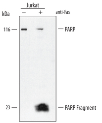
- Detection of Human PARP by Western Blot. Western blot shows lysates of Jurkat human acute T cell leukemia cell line untreated (-) or treated (+) with 200 ng/mL anti-Fas for 24 hours. PVDF membrane was probed with 0.4 µg/mL of Goat Anti-Human/Mouse PARP Affinity-purified Polyclonal Antibody (Catalog # AF-600-NA) followed by HRP-conjugated Anti-Goat IgG Secondary Antibody (Catalog # HAF109). A specific band was detected for PARP at approximately 116 kDa (as indicated). This experiment was conducted under reducing conditions and using Immunoblot Buffer Group 2.
- Submitted by
- R&D Systems (provider)
- Main image
- Experimental details
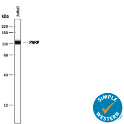
- Detection of Human PARP by Simple WesternTM. Simple Western lane view shows lysates of Jurkat human acute T cell leukemia cell line, loaded at 0.2 mg/mL. A specific band was detected for PARP at approximately 122 kDa (as indicated) using 5 µg/mL of Goat Anti-Human/Mouse PARP Antigen Affinity-purified Polyclonal Antibody (Catalog # AF-600-NA) followed by 1:50 dilution of HRP-conjugated Anti-Goat IgG Secondary Antibody (Catalog # HAF109). This experiment was conducted under reducing conditions and using the 12-230 kDa separation system.

- Submitted by
- R&D Systems (provider)
- Main image
- Experimental details
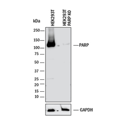
- Western Blot Shows Human PARP Specificity by Using Knockout Cell Line. Western blot shows lysates of HEK293T human embryonic kidney parental cell line and PARP knockout HEK293T cell line (KO). PVDF membrane was probed with 0.4 µg/mL of Goat Anti-Human/Mouse PARP Antigen Affinity-purified Polyclonal Antibody (Catalog # AF-600-NA) followed by HRP-conjugated Anti-Goat IgG Secondary Antibody (Catalog # HAF017). A specific band was detected for PARP at approximately 120 kDa (as indicated) in the parental HEK293T cell line, but is not detectable in knockout HEK293Tcell line. GAPDH (Catalog # AF5718) is shown as a loading control. This experiment was conducted under reducing conditions and using Immunoblot Buffer Group 1.

Supportive validation
- Submitted by
- R&D Systems (provider)
- Main image
- Experimental details
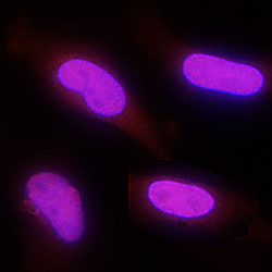
- PARP in HeLa Human Cell Line. PARP was detected in immersion fixed HeLa human cervical epithelial carcinoma cell line using Goat Anti-Human/Mouse PARP Antigen Affinity-purified Polyclonal Antibody (Catalog # AF-600-NA) at 1 µg/mL for 3 hours at room temperature. Cells were stained using the NorthernLights™ 557-conjugated Anti-Goat IgG Secondary Antibody (red; Catalog # NL001) and counterstained with DAPI (blue). Specific staining was localized to nuclei. View our protocol for Fluorescent ICC Staining of Cells on Coverslips.

Supportive validation
- Submitted by
- R&D Systems (provider)
- Main image

- Experimental details
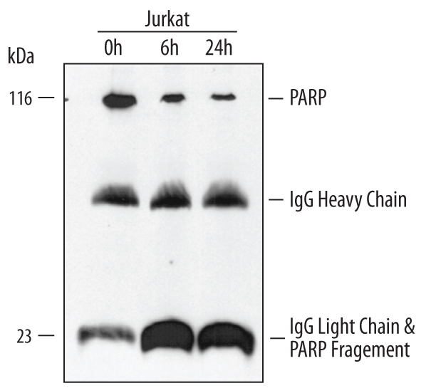
- Immunoprecipitation of Human PARP. Jurkat human acute T cell leukemia cell line was treated with apoptosis inducer anti-Fas for the indicated times. PARP was immunoprecipitated from cell lysates (1 - 2 x 10⁶ cells) following incubation with 5 µg Goat Anti-Human/Mouse PARP Antigen Affinity-purified Polyclonal Antibody (Catalog # AF-600-NA) for overnight at 4 °C. PARP-antibody complexes were absorbed using Protein G expressing Staph cells (Sigma). Immunoprecipitated PARP was detected by Western blot using 0.4 µg/mL Goat Anti-Human/Mouse PARP Antigen Affinity-purified Polyclonal Antibody (Catalog # AF-600-NA). View our recommended buffer recipes for immunoprecipitation.