 Explore
 Validate
 Learn
Learn Western blot
Antibody data
- Antibody Data
- Antigen structure
- References [4]
- Comments [0]
- Validations
- Western blot [2]
Submit
Validation data
Reference
Comment
Report error
- Product number
- MAB600 - Provider product page

- Provider
- R&D Systems
- Product name
- Human/Mouse PARP Antibody
- Antibody type
- Monoclonal
- Description
- Protein A or G purified from hybridoma culture supernatant. Detects human and mouse PARP in Western blots.
- Reactivity
- Human, Mouse
- Host
- Rat
- Conjugate
- Unconjugated
- Antigen sequence
NP_031441- Isotype
- IgG
- Antibody clone number
- 53015
- Vial size
- 500 ug
- Storage
- Use a manual defrost freezer and avoid repeated freeze-thaw cycles. 12 months from date of receipt, -20 to -70 °C as supplied. 1 month, 2 to 8 °C under sterile conditions after reconstitution. 6 months, -20 to -70 °C under sterile conditions after reconstitution.
Submitted references RNA stability regulates human T cell leukemia virus type 1 gene expression in chronically-infected CD4 T cells.
Distinct promoters mediate constitutive and inducible Bcl-XL expression in malignant lymphocytes.
Conditional deletion of Nbs1 in murine cells reveals its role in branching repair pathways of DNA double-strand breaks.
Transcriptional repressor activating transcription factor 3 protects human umbilical vein endothelial cells from tumor necrosis factor-alpha-induced apoptosis through down-regulation of p53 transcription.
Lin HC, Simon PJ, Ysla RM, Zeichner SL, Brewer G, Rabson AB
Virology 2017 Aug;508:7-17
Virology 2017 Aug;508:7-17
Distinct promoters mediate constitutive and inducible Bcl-XL expression in malignant lymphocytes.
Habens F, Lapham AS, Dallman CL, Pickering BM, Michels J, Marcusson EG, Johnson PW, Packham G
Oncogene 2007 Mar 22;26(13):1910-9
Oncogene 2007 Mar 22;26(13):1910-9
Conditional deletion of Nbs1 in murine cells reveals its role in branching repair pathways of DNA double-strand breaks.
Yang YG, Saidi A, Frappart PO, Min W, Barrucand C, Dumon-Jones V, Michelon J, Herceg Z, Wang ZQ
The EMBO journal 2006 Nov 29;25(23):5527-38
The EMBO journal 2006 Nov 29;25(23):5527-38
Transcriptional repressor activating transcription factor 3 protects human umbilical vein endothelial cells from tumor necrosis factor-alpha-induced apoptosis through down-regulation of p53 transcription.
Kawauchi J, Zhang C, Nobori K, Hashimoto Y, Adachi MT, Noda A, Sunamori M, Kitajima S
The Journal of biological chemistry 2002 Oct 11;277(41):39025-34
The Journal of biological chemistry 2002 Oct 11;277(41):39025-34
No comments: Submit comment
Supportive validation
- Submitted by
- R&D Systems (provider)
- Main image
- Experimental details
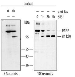
- Detection of Human PARP by Western Blot. Western blot shows lysates of Jurkat human acute T cell leukemia cell line untreated (-) or treated with anti-Fas and 1 µM staurosporine (STS) for 0 to 4 hours (h; as indicated). PVDF membrane was probed with 0.6 µg/mL of Rat Anti-Human/Mouse PARP Monoclonal Antibody (Catalog # MAB600) followed by HRP-conjugated Anti-Rat IgG Secondary Antibody (Catalog # HAF005). Specific bands were detected for PARP at approximately 116 kDa and the 84 kDa product of PARP cleavage (as indicated). This experiment was conducted under reducing conditions and using Immunoblot Buffer Group 2.

- Submitted by
- R&D Systems (provider)
- Main image

- Experimental details
- Detection of Mouse PARP by Western Blot. Western blot shows lysates of NIH-3T3 mouse embryonic fibroblast cell line treated with 1 µM staurosporine (STS) for 0 to 4 hours (h; as indicated). PVDF membrane was probed with 0.6 µg/mL of Rat Anti-Human/Mouse PARP Monoclonal Antibody (Catalog # MAB600) followed by HRP-conjugated Anti-Rat IgG Secondary Antibody (Catalog # HAF005). Specific bands were detected for PARP at approximately 116 kDa and the 60-70 kDa bands generated with staurosporine treatment (as indicated). This experiment was conducted under reducing conditions and using Immunoblot Buffer Group 2.
