 Explore
 Validate
 Learn
Learn Western blot
Antibody data
- Antibody Data
- Antigen structure
- References [3]
- Comments [0]
- Validations
- Western blot [1]
- Immunohistochemistry [1]
- Other assay [2]
Submit
Validation data
Reference
Comment
Report error
- Product number
- 44-1026G - Provider product page

- Provider
- Invitrogen Antibodies
- Product name
- Phospho-Paxillin (Ser178) Polyclonal Antibody
- Antibody type
- Polyclonal
- Antigen
- Synthetic peptide
- Reactivity
- Human
- Host
- Rabbit
- Isotype
- IgG
- Vial size
- 100 µL
- Storage
- -20°C
Submitted references Inhibition of the invasion and migration of renal carcinoma 786‑o‑si3 cells in vitro and in vivo by Koelreuteria formosana extract.
Regulation of neurite growth by inorganic pyrophosphatase 1 via JNK dephosphorylation.
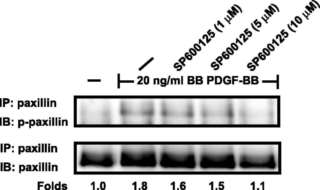
c-Jun N-terminal kinase is necessary for platelet-derived growth factor-mediated chemotaxis in primary fibroblasts.
Lin CY, Chen PN, Hsu LS, Kuo DY, Chu SC, Hsieh YS
Molecular medicine reports 2014 Dec;10(6):3334-42
Molecular medicine reports 2014 Dec;10(6):3334-42
Regulation of neurite growth by inorganic pyrophosphatase 1 via JNK dephosphorylation.
Tezuka Y, Okada M, Tada Y, Yamauchi J, Nishigori H, Sanbe A
PloS one 2013;8(4):e61649
PloS one 2013;8(4):e61649
c-Jun N-terminal kinase is necessary for platelet-derived growth factor-mediated chemotaxis in primary fibroblasts.
Amagasaki K, Kaneto H, Heldin CH, Lennartsson J
The Journal of biological chemistry 2006 Aug 4;281(31):22173-22179
The Journal of biological chemistry 2006 Aug 4;281(31):22173-22179
No comments: Submit comment
Supportive validation
- Submitted by
- Invitrogen Antibodies (provider)
- Main image
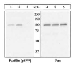
- Experimental details
- Lysates from untreated EGFP-Paxillin β-transfected Hek293 cells (1, 4), lysates from EGFP-Paxillin β-transfected Hek-293 cells treated with EGF (2, 5), and lysates from EGFP-Paxillin S178A mutant tranfected Hek-293 treated with EGF (3, 6), were resolved on a 10% polyacrylamide gel and transferred to PVDF. Membranes were blocked with a 5% milk-TBST buffer for one hour at room temperature, and then were incubated with the Paxillin (pS178) antibody (1-3) or anti-Paxillin pan antibody (4-6) for two hours at room temperature in a 1% milk-TBST buffer. After washing, membranes were incubated with goat F (ab')2 anti-rabbit IgG HRP conjugate, and bands were detected using the Pierce SuperSignal™ method.
Supportive validation
- Submitted by
- Invitrogen Antibodies (provider)
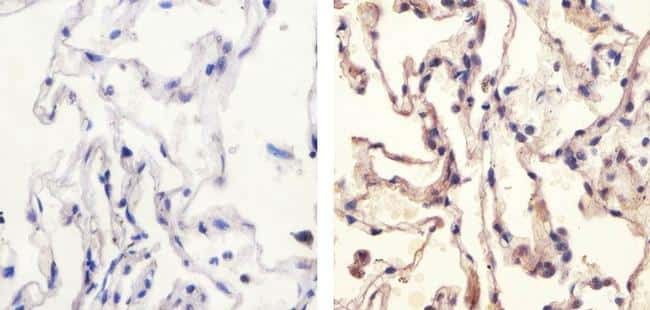
- Main image

- Experimental details
- Immunohistochemistry analysis of Phospho-Paxillin pSer178 showing staining in the cytoplasm of paraffin-embedded human lung tissue (right) compared to a negative control without primary antibody (left). To expose target proteins, antigen retrieval was performed using 10mM sodium citrate (pH 6.0), microwaved for 8-15 min. Following antigen retrieval, tissues were blocked in 3% H2O2-methanol for 15 min at room temperature, washed with ddH2O and PBS, and then probed with a Anti- Phospho-Paxillin pSer178 Polyclonal Antibody (Product # 44-1026G) diluted in 3% BSA-PBS at a dilution of 1:20 overnight at 4°C in a humidified chamber. Tissues were washed extensively in PBST and detection was performed using an HRP-conjugated secondary antibody followed by colorimetric detection using a DAB kit. Tissues were counterstained with hematoxylin and dehydrated with ethanol and xylene to prep for mounting.
Supportive validation
- Submitted by
- Invitrogen Antibodies (provider)
- Main image

- Experimental details
- NULL
- Submitted by
- Invitrogen Antibodies (provider)
- Main image
- Experimental details
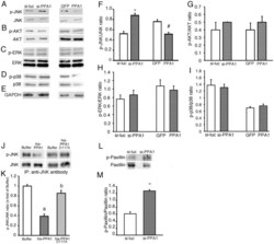
- Figure 4 Phosphorylated protein kinase levels in N1E115 cells. (A-E) Representative Western blots using anti-phospho-JNK and JNK (A), phospho-AKT and AKT (B), phospho-ERK and ERK (C), phospho p38 and p38 (D) and GAPDH (E). N1E115 cells were treated with si-RNA targeted to mouse PPA1 (si-PPA1) or treated with adenovirus containing mouse PPA1 (PPA1). Si-RNA targeted to luciferase (si-luc) was used as a si-PPA1 control and adenovirus containing GFP (GFP) was used as a control for adenovirus containing mouse PPA1. (F-I) Quantitative analysis of the phosphorylated JNK/JNK ratio (F), pAKT/AKT ratio (G), pERK/ERK ratio (H) and p-p38/p38 ratio (I) are shown. (J and K) Direct effects of recombinant PPA1 (his-PPA1) or PPA1 D117A (his-PPA1 D117A) proteins on the phosphorylated JNK level immunoprecipitated from N1E115 cells. As a control, buffer without the recombinant protein was added (buffer). (n = 5) Representative Western blot using anti-phospho-JNK and JNK (J) and quantitative analysis of the phosphorylated JNK/JNK ratio (K) (n = 5). (L and M) Phosphorylated paxillin level in N1E115 cells. Representative Western blot using anti-phospho-paxillin and paxillin (L) and quantitative analysis of the phosphorylated paxillin/paxillin ratio (M) (n = 5). *p
