 Explore
 Validate
 Learn
Learn Western blot
 Immunoprecipitation
Antibody data
- Antibody Data
- Antigen structure
- References [0]
- Comments [0]
- Validations
- Western blot [8]
- Immunocytochemistry [1]
- Immunohistochemistry [3]
- Other assay [1]
Submit
Validation data
Reference
Comment
Report error
- Product number
- PA5-34910 - Provider product page

- Provider
- Invitrogen Antibodies
- Product name
- Paxillin Polyclonal Antibody
- Antibody type
- Polyclonal
- Antigen
- Recombinant protein fragment
- Description
- Recommended positive controls: 293T, A431, HeLa, HepG2, rat brain, GFP-human Paxillin-transfected 293T, MDA-MB-231. Predicted reactivity: Mouse (94%), Chicken (82%). Store product as a concentrated solution. Centrifuge briefly prior to opening the vial.
- Reactivity
- Human, Mouse, Rat
- Host
- Rabbit
- Isotype
- IgG
- Vial size
- 100 µL
- Concentration
- 0.39 mg/mL
- Storage
- Store at 4°C short term. For long term storage, store at -20°C, avoiding freeze/thaw cycles.
No comments: Submit comment
Supportive validation
- Submitted by
- Invitrogen Antibodies (provider)
- Main image
- Experimental details
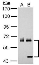
- Western blot analysis of Paxillin using A) 30 µg K562 whole cell lysate and B) 30 µg THP-1 whole cell lysate. Samples were loaded onto a 7.5% SDS-PAGE gel and probed with a Paxillin polyclonal antibody (Product # PA5-34910) at a dilution of 1:1000.

- Submitted by
- Invitrogen Antibodies (provider)
- Main image

- Experimental details
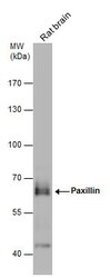
- Western blot analysis of Paxillin in Rat tissue extract (50 µg). Samples was separated by 7.5% SDS-PAGE and the membrane was probed with Paxillin Polyclonal antibody (Product # PA5-34910) at a dilution of 1:1000.
- Submitted by
- Invitrogen Antibodies (provider)
- Main image

- Experimental details
- Western Blot analysis of Paxillin was performed by separating 30 µg of various whole cell extracts by 7.5% SDS-PAGE. Proteins were transferred to a membrane and probed with a Paxillin Polyclonal Antibody (Product # PA5-34910) at a dilution of 1:5000 and a HRP-conjugated anti-rabbit IgG secondary antibody.

- Submitted by
- Invitrogen Antibodies (provider)
- Main image
- Experimental details
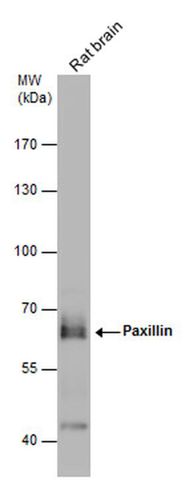
- Western Blot using Paxillin Polyclonal Antibody (Product # PA5-34910). Rat tissue extract (50 µg) was separated by 7.5% SDS-PAGE, and the membrane was blotted with Paxillin Polyclonal Antibody (Product # PA5-34910) diluted at 1:1,000. The HRP-conjugated anti-rabbit IgG antibody was used to detect the primary antibody.

- Submitted by
- Invitrogen Antibodies (provider)
- Main image

- Experimental details
- Paxillin Polyclonal Antibody detects Paxillin protein by western blot analysis. A. 1 µg 293T whole cell extract. B. 1 µg whole cell extract of GFP-human Paxillin-transfected 293T cells.7.5% SDS-PAGE. Paxillin Polyclonal Antibody (Product # PA5-34910) dilution: 1:10,000. The HRP-conjugated anti-rabbit IgG antibody was used to detect the primary antibody.

- Submitted by
- Invitrogen Antibodies (provider)
- Main image

- Experimental details
- Western Blot using Paxillin Polyclonal Antibody (Product # PA5-34910). Various whole cell extracts (30 µg) were separated by 7.5% SDS-PAGE, and the membrane was blotted with Paxillin Polyclonal Antibody (Product # PA5-34910) diluted at 1:5,000. The HRP-conjugated anti-rabbit IgG antibody was used to detect the primary antibody.

- Submitted by
- Invitrogen Antibodies (provider)
- Main image

- Experimental details
- Knockout of Paxillin was achieved by CRISPR-Cas9 genome editing using LentiArray™ Lentiviral sgRNA (Product # A32042, Assay ID CRISPR1071991_LV) and LentiArray Cas9 Lentivirus (Product # A32064). Western blot analysis of Paxillin was performed by loading 30 µg of HeLa Wild Type (Lane 1), HeLa Cas9 (Lane 2) andHeLa Paxillin KO (Lane 3) whole cell extracts. The samples were electrophoresed using NuPAGE™ Novex™ 4-12% Bis-Tris Protein Gel (Product # NP0322BOX). Resolved proteins were then transferred onto a nitrocellulose membrane (Product # IB23001) by iBlot® 2 Dry Blotting System (Product # IB21001). The blot was probed with Anti-Paxillin Polyclonal Antibody (Product # PA5-34910, 1:7,000 dilution) and Goat anti-Rabbit IgG (H+L) Superclonal™ Recombinant Secondary Antibody, HRP (Product # A27036, 1:10,000 dilution) using the iBright FL 1000 (Product # A32752). Chemiluminescent detection was performed using Novex® ECL Chemiluminescent Substrate Reagent Kit (Product # WP20005). Loss of signal upon CRISPR mediated knockout (KO) using the LentiArray™ CRISPR product line confirms that antibody is specific to Paxillin. An uncharacterized band was observed in all the samples at 32 kDa.

- Submitted by
- Invitrogen Antibodies (provider)
- Main image

- Experimental details
- Western blot was performed using Anti-Paxillin Polyclonal Antibody(Product # PA5-34910) and a 64kDa band corresponding to Paxillin was observed across all tested cell lines and tissues, except MCF7. Whole cell extracts (30 µg lysate) of A-431 (Lane 1), HeLa (Lane 2), COS-7 (Lane 3), PC-12 (Lane 4), Caco-2 (Lane 5), MCF7 (Lane 6), T-47D (Lane 7), Mouse Lung (Lane 8), Rat Lung (Lane 9), NIH/3T3 (Lane 10) were electrophoresed using NuPAGE™ 10% Bis-Tris Protein Gel (Product # NP0302BOX). Resolved proteins were then transferred onto a Nitrocellulose membrane (Product # IB23001) by iBlot® 2 Dry Blotting System (Product # IB21001). The blot was probed with the primary antibody (1:5000) and detected by chemiluminescence with Goat anti-Rabbit IgG (H+L) Superclonal™ Recombinant Secondary Antibody, HRP (Product # A27036,1:4000) using the iBright FL 1000 (Product # A32752). Chemiluminescent detection was performed using Novex® ECL Chemiluminescent Substrate Reagent Kit (Product # WP20005).

Supportive validation
- Submitted by
- Invitrogen Antibodies (provider)
- Main image

- Experimental details
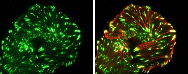
- Immunocytochemistry-Immunofluorescence analysis of Paxillin was performed in MDA-MB-231 cells fixed in 4% paraformaldehyde at RT for 15 min. Green: Paxillin Polyclonal Antibody (Product # PA5-34910) diluted at 1:100. Red: phalloidin staining.
Supportive validation
- Submitted by
- Invitrogen Antibodies (provider)
- Main image

- Experimental details
- Immunohistochemistry (Paraffin) analysis of Paxillin was performed in paraffin-embedded mouse testis tissue using Paxillin Polyclonal Antibody (Product # PA5-34910) at a dilution of 1:500. Antigen Retrieval: Citrate buffer, pH 6.0, 15 min.
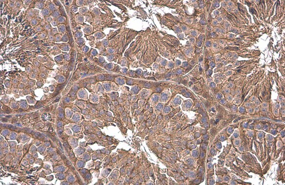
- Submitted by
- Invitrogen Antibodies (provider)
- Main image

- Experimental details
- Immunohistochemistry (Paraffin) analysis of Paxillin was performed in paraffin-embedded rat testis tissue using Paxillin Polyclonal Antibody (Product # PA5-34910) at a dilution of 1:500. Antigen Retrieval: Citrate buffer, pH 6.0, 15 min.

- Submitted by
- Invitrogen Antibodies (provider)
- Main image

- Experimental details
- Paxillin Polyclonal Antibody detects Paxillin protein at cell membrane and cytoplasm by immunohistochemical analysis. Sample: Paraffin-embedded mouse testis. Paxillin stained by Paxillin Polyclonal Antibody (Product # PA5-34910) diluted at 1:500. Antigen Retrieval: Citrate buffer, pH 6.0, 15 min.

Supportive validation
- Submitted by
- Invitrogen Antibodies (provider)
- Main image
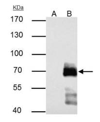
- Experimental details
- Paxillin Polyclonal Antibody immunoprecipitates Paxillin protein in IP experiments. IP samples: HeLa whole cell extract. A. Control with 2 µg of preimmune Rabbit IgG. B. Immunoprecipitation of Paxillin protein by 2 µg Paxillin Polyclonal Antibody (Product # PA5-34910). 7.5 % SDS-PAGE. The immunoprecipitated Paxillin protein was detected by Paxillin Polyclonal Antibody (Product # PA5-34910) diluted at 1:1,0000.
