 Explore
 Validate
 Learn
Learn Western blot
 Immunohistochemistry
Antibody data
- Antibody Data
- Antigen structure
- References [34]
- Comments [0]
- Validations
- Western blot [1]
- Immunocytochemistry [2]
- Other assay [6]
Submit
Validation data
Reference
Comment
Report error
- Product number
- 44-720G - Provider product page

- Provider
- Invitrogen Antibodies
- Product name
- Phospho-Paxillin (Tyr31) Polyclonal Antibody
- Antibody type
- Polyclonal
- Antigen
- Synthetic peptide
- Reactivity
- Human, Mouse
- Host
- Rabbit
- Isotype
- IgG
- Vial size
- 100 µL
- Storage
- -20°C
Submitted references Matrix stiffness modulates tip cell formation through the p-PXN-Rac1-YAP signaling axis.
ARHGEF9 regulates melanoma morphogenesis in environments with diverse geometry and elasticity by promoting filopodial-driven adhesion.
Heterochromatin-Driven Nuclear Softening Protects the Genome against Mechanical Stress-Induced Damage.
Basement Membrane Regulates Fibronectin Organization Using Sliding Focal Adhesions Driven by a Contractile Winch.
RGD-functionalized supported lipid bilayers modulate pre-osteoblast adherence and promote osteogenic differentiation.
Phospholipid phosphatase related 1 (PLPPR1) increases cell adhesion through modulation of Rac1 activity.
Coupling of β(2) integrins to actin by a mechanosensitive molecular clutch drives complement receptor-mediated phagocytosis.
Plk1 Mediates Paxillin Phosphorylation (Ser-272), Centrosome Maturation, and Airway Smooth Muscle Layer Thickening in Allergic Asthma.
Talin-mediated force transmission and talin rod domain unfolding independently regulate adhesion signaling.
Enhancement of malignant properties of human glioma cells by ganglioside GD3/GD2.
Nicotinic acid impairs assembly of leading edge in glioma cells.
15-Deoxy-Δ(12,14)-prostaglandin J(2) inhibits migration of human thyroid carcinoma cells by disrupting focal adhesion complex and adherens junction.
The Integrin-Mediated ILK-Parvin-αPix Signaling Axis Controls Differentiation in Mammary Epithelial Cells.
RhoB controls endothelial barrier recovery by inhibiting Rac1 trafficking to the cell border.
Talin tension sensor reveals novel features of focal adhesion force transmission and mechanosensitivity.
Localized LoxL3-Dependent Fibronectin Oxidation Regulates Myofiber Stretch and Integrin-Mediated Adhesion.
Kindlin-2 cooperates with talin to activate integrins and induces cell spreading by directly binding paxillin.
FMN2 Makes Perinuclear Actin to Protect Nuclei during Confined Migration and Promote Metastasis.
Focal adhesion kinase-dependent focal adhesion recruitment of SH2 domains directs SRC into focal adhesions to regulate cell adhesion and migration.
Dissecting motility signaling through activation of specific Src-effector complexes.
Governing epidermal homeostasis by coupling cell-cell adhesion to integrin and growth factor signaling, proliferation, and apoptosis.
Biophysical stimulation induces demyelination via an integrin-dependent mechanism.
Knock-in mutation reveals an essential role for focal adhesion kinase activity in blood vessel morphogenesis and cell motility-polarity but not cell proliferation.
Apoptosis commitment and activation of mitochondrial Bax during anoikis is regulated by p38MAPK.
Src inhibitors in early breast cancer: a methodology, feasibility and variability study.
Paxillin-Y118 phosphorylation contributes to the control of Src-induced anchorage-independent growth by FAK and adhesion.
Paxillin phosphorylation, actin polymerization, noise temperature, and the sustained phase of swine carotid artery contraction.
Inhibition of cell migration by autophosphorylated mammalian sterile 20-like kinase 3 (MST3) involves paxillin and protein-tyrosine phosphatase-PEST.
Glycogen synthase kinase 3- and extracellular signal-regulated kinase-dependent phosphorylation of paxillin regulates cytoskeletal rearrangement.
Mislocalization or reduced expression of Arf GTPase-activating protein ASAP1 inhibits cell spreading and migration by influencing Arf1 GTPase cycling.
p130cas but not paxillin is essential for Caco-2 intestinal epithelial cell spreading and migration on collagen IV.
Aberrant activation of focal adhesion proteins mediates fibrillar amyloid beta-induced neuronal dystrophy.
Aberrant activation of focal adhesion proteins mediates fibrillar amyloid beta-induced neuronal dystrophy.
Tyrosine phosphorylation of paxillin alpha is involved in temporospatial regulation of paxillin-containing focal adhesion formation and F-actin organization in motile cells.
Guo Y, Mei F, Huang Y, Ma S, Wei Y, Zhang X, Xu M, He Y, Heng BC, Chen L, Deng X
Bioactive materials 2022 Jan;7:364-376
Bioactive materials 2022 Jan;7:364-376
ARHGEF9 regulates melanoma morphogenesis in environments with diverse geometry and elasticity by promoting filopodial-driven adhesion.
Bousgouni V, Inge O, Robertson D, Jones I, Clatworthy I, Bakal C
iScience 2022 Aug 19;25(8):104795
iScience 2022 Aug 19;25(8):104795
Heterochromatin-Driven Nuclear Softening Protects the Genome against Mechanical Stress-Induced Damage.
Nava MM, Miroshnikova YA, Biggs LC, Whitefield DB, Metge F, Boucas J, Vihinen H, Jokitalo E, Li X, García Arcos JM, Hoffmann B, Merkel R, Niessen CM, Dahl KN, Wickström SA
Cell 2020 May 14;181(4):800-817.e22
Cell 2020 May 14;181(4):800-817.e22
Basement Membrane Regulates Fibronectin Organization Using Sliding Focal Adhesions Driven by a Contractile Winch.
Lu J, Doyle AD, Shinsato Y, Wang S, Bodendorfer MA, Zheng M, Yamada KM
Developmental cell 2020 Mar 9;52(5):631-646.e4
Developmental cell 2020 Mar 9;52(5):631-646.e4
RGD-functionalized supported lipid bilayers modulate pre-osteoblast adherence and promote osteogenic differentiation.
Verstappen JFM, Jin J, Koçer G, Haroon M, Jonkheijm P, Bakker AD, Klein-Nulend J, Jaspers RT
Journal of biomedical materials research. Part A 2020 Apr;108(4):923-937
Journal of biomedical materials research. Part A 2020 Apr;108(4):923-937
Phospholipid phosphatase related 1 (PLPPR1) increases cell adhesion through modulation of Rac1 activity.
Tilve S, Iweka CA, Bao J, Hawken N, Mencio CP, Geller HM
Experimental cell research 2020 Apr 15;389(2):111911
Experimental cell research 2020 Apr 15;389(2):111911
Coupling of β(2) integrins to actin by a mechanosensitive molecular clutch drives complement receptor-mediated phagocytosis.
Jaumouillé V, Cartagena-Rivera AX, Waterman CM
Nature cell biology 2019 Nov;21(11):1357-1369
Nature cell biology 2019 Nov;21(11):1357-1369
Plk1 Mediates Paxillin Phosphorylation (Ser-272), Centrosome Maturation, and Airway Smooth Muscle Layer Thickening in Allergic Asthma.
Rezey AC, Gerlach BD, Wang R, Liao G, Tang DD
Scientific reports 2019 May 17;9(1):7555
Scientific reports 2019 May 17;9(1):7555
Talin-mediated force transmission and talin rod domain unfolding independently regulate adhesion signaling.
Rahikainen R, Öhman T, Turkki P, Varjosalo M, Hytönen VP
Journal of cell science 2019 Apr 3;132(7)
Journal of cell science 2019 Apr 3;132(7)
Enhancement of malignant properties of human glioma cells by ganglioside GD3/GD2.
Iwasawa T, Zhang P, Ohkawa Y, Momota H, Wakabayashi T, Ohmi Y, Bhuiyan RH, Furukawa K, Furukawa K
International journal of oncology 2018 Apr;52(4):1255-1266
International journal of oncology 2018 Apr;52(4):1255-1266
Nicotinic acid impairs assembly of leading edge in glioma cells.
Yang X, Mei S, Niu H, Li J
Oncology reports 2017 Aug;38(2):829-836
Oncology reports 2017 Aug;38(2):829-836
15-Deoxy-Δ(12,14)-prostaglandin J(2) inhibits migration of human thyroid carcinoma cells by disrupting focal adhesion complex and adherens junction.
Wu YC, Jhao YT, Cheng YC, Chen Y
Oncology letters 2017 Apr;13(4):2569-2576
Oncology letters 2017 Apr;13(4):2569-2576
The Integrin-Mediated ILK-Parvin-αPix Signaling Axis Controls Differentiation in Mammary Epithelial Cells.
Rooney N, Wang P, Brennan K, Gilmore AP, Streuli CH
Journal of cellular physiology 2016 Nov;231(11):2408-17
Journal of cellular physiology 2016 Nov;231(11):2408-17
RhoB controls endothelial barrier recovery by inhibiting Rac1 trafficking to the cell border.
Marcos-Ramiro B, García-Weber D, Barroso S, Feito J, Ortega MC, Cernuda-Morollón E, Reglero-Real N, Fernández-Martín L, Durán MC, Alonso MA, Correas I, Cox S, Ridley AJ, Millán J
The Journal of cell biology 2016 May 9;213(3):385-402
The Journal of cell biology 2016 May 9;213(3):385-402
Talin tension sensor reveals novel features of focal adhesion force transmission and mechanosensitivity.
Kumar A, Ouyang M, Van den Dries K, McGhee EJ, Tanaka K, Anderson MD, Groisman A, Goult BT, Anderson KI, Schwartz MA
The Journal of cell biology 2016 May 9;213(3):371-83
The Journal of cell biology 2016 May 9;213(3):371-83
Localized LoxL3-Dependent Fibronectin Oxidation Regulates Myofiber Stretch and Integrin-Mediated Adhesion.
Kraft-Sheleg O, Zaffryar-Eilot S, Genin O, Yaseen W, Soueid-Baumgarten S, Kessler O, Smolkin T, Akiri G, Neufeld G, Cinnamon Y, Hasson P
Developmental cell 2016 Mar 7;36(5):550-61
Developmental cell 2016 Mar 7;36(5):550-61
Kindlin-2 cooperates with talin to activate integrins and induces cell spreading by directly binding paxillin.
Theodosiou M, Widmaier M, Böttcher RT, Rognoni E, Veelders M, Bharadwaj M, Lambacher A, Austen K, Müller DJ, Zent R, Fässler R
eLife 2016 Jan 27;5:e10130
eLife 2016 Jan 27;5:e10130
FMN2 Makes Perinuclear Actin to Protect Nuclei during Confined Migration and Promote Metastasis.
Skau CT, Fischer RS, Gurel P, Thiam HR, Tubbs A, Baird MA, Davidson MW, Piel M, Alushin GM, Nussenzweig A, Steeg PS, Waterman CM
Cell 2016 Dec 1;167(6):1571-1585.e18
Cell 2016 Dec 1;167(6):1571-1585.e18
Focal adhesion kinase-dependent focal adhesion recruitment of SH2 domains directs SRC into focal adhesions to regulate cell adhesion and migration.
Wu JC, Chen YC, Kuo CT, Wenshin Yu H, Chen YQ, Chiou A, Kuo JC
Scientific reports 2015 Dec 18;5:18476
Scientific reports 2015 Dec 18;5:18476
Dissecting motility signaling through activation of specific Src-effector complexes.
Karginov AV, Tsygankov D, Berginski M, Chu PH, Trudeau ED, Yi JJ, Gomez S, Elston TC, Hahn KM
Nature chemical biology 2014 Apr;10(4):286-90
Nature chemical biology 2014 Apr;10(4):286-90
Governing epidermal homeostasis by coupling cell-cell adhesion to integrin and growth factor signaling, proliferation, and apoptosis.
Livshits G, Kobielak A, Fuchs E
Proceedings of the National Academy of Sciences of the United States of America 2012 Mar 27;109(13):4886-91
Proceedings of the National Academy of Sciences of the United States of America 2012 Mar 27;109(13):4886-91
Biophysical stimulation induces demyelination via an integrin-dependent mechanism.
Lin MY, Frieboes LS, Forootan M, Palispis WA, Mozaffar T, Jafari M, Steward O, Gall CM, Gupta R
Annals of neurology 2012 Jul;72(1):112-23
Annals of neurology 2012 Jul;72(1):112-23
Knock-in mutation reveals an essential role for focal adhesion kinase activity in blood vessel morphogenesis and cell motility-polarity but not cell proliferation.
Lim ST, Chen XL, Tomar A, Miller NL, Yoo J, Schlaepfer DD
The Journal of biological chemistry 2010 Jul 9;285(28):21526-36
The Journal of biological chemistry 2010 Jul 9;285(28):21526-36
Apoptosis commitment and activation of mitochondrial Bax during anoikis is regulated by p38MAPK.
Owens TW, Valentijn AJ, Upton JP, Keeble J, Zhang L, Lindsay J, Zouq NK, Gilmore AP
Cell death and differentiation 2009 Nov;16(11):1551-62
Cell death and differentiation 2009 Nov;16(11):1551-62
Src inhibitors in early breast cancer: a methodology, feasibility and variability study.
Jones RJ, Young O, Renshaw L, Jacobs V, Fennell M, Marshall A, Green TP, Elvin P, Womack C, Clack G, Dixon JM
Breast cancer research and treatment 2009 Mar;114(2):211-21
Breast cancer research and treatment 2009 Mar;114(2):211-21
Paxillin-Y118 phosphorylation contributes to the control of Src-induced anchorage-independent growth by FAK and adhesion.
Sachdev S, Bu Y, Gelman IH
BMC cancer 2009 Jan 12;9:12
BMC cancer 2009 Jan 12;9:12
Paxillin phosphorylation, actin polymerization, noise temperature, and the sustained phase of swine carotid artery contraction.
Rembold CM, Tejani AD, Ripley ML, Han S
American journal of physiology. Cell physiology 2007 Sep;293(3):C993-1002
American journal of physiology. Cell physiology 2007 Sep;293(3):C993-1002
Inhibition of cell migration by autophosphorylated mammalian sterile 20-like kinase 3 (MST3) involves paxillin and protein-tyrosine phosphatase-PEST.
Lu TJ, Lai WY, Huang CY, Hsieh WJ, Yu JS, Hsieh YJ, Chang WT, Leu TH, Chang WC, Chuang WJ, Tang MJ, Chen TY, Lu TL, Lai MD
The Journal of biological chemistry 2006 Dec 15;281(50):38405-17
The Journal of biological chemistry 2006 Dec 15;281(50):38405-17
Glycogen synthase kinase 3- and extracellular signal-regulated kinase-dependent phosphorylation of paxillin regulates cytoskeletal rearrangement.
Cai X, Li M, Vrana J, Schaller MD
Molecular and cellular biology 2006 Apr;26(7):2857-68
Molecular and cellular biology 2006 Apr;26(7):2857-68
Mislocalization or reduced expression of Arf GTPase-activating protein ASAP1 inhibits cell spreading and migration by influencing Arf1 GTPase cycling.
Liu Y, Yerushalmi GM, Grigera PR, Parsons JT
The Journal of biological chemistry 2005 Mar 11;280(10):8884-92
The Journal of biological chemistry 2005 Mar 11;280(10):8884-92
p130cas but not paxillin is essential for Caco-2 intestinal epithelial cell spreading and migration on collagen IV.
Sanders MA, Basson MD
The Journal of biological chemistry 2005 Jun 24;280(25):23516-22
The Journal of biological chemistry 2005 Jun 24;280(25):23516-22
Aberrant activation of focal adhesion proteins mediates fibrillar amyloid beta-induced neuronal dystrophy.
Grace EA, Busciglio J
The Journal of neuroscience : the official journal of the Society for Neuroscience 2003 Jan 15;23(2):493-502
The Journal of neuroscience : the official journal of the Society for Neuroscience 2003 Jan 15;23(2):493-502
Tyrosine phosphorylation of paxillin alpha is involved in temporospatial regulation of paxillin-containing focal adhesion formation and F-actin organization in motile cells.
Nakamura K, Yano H, Uchida H, Hashimoto S, Schaefer E, Sabe H
The Journal of biological chemistry 2000 Sep 1;275(35):27155-64
The Journal of biological chemistry 2000 Sep 1;275(35):27155-64
No comments: Submit comment
Supportive validation
- Submitted by
- Invitrogen Antibodies (provider)
- Main image

- Experimental details
- Western blot analysis was performed on whole cell extracts (20 µg lysate) of A431 (lane 1), A431 treated for 1 Hr with 100 uM of Pervanadate (lane 2), HeLa (lane 3), HeLa treated for 20 minutes with 50 ng/mL of TNF alpha (lane 4), NIH/3T3 (lane 5), NIH/3T3 treated for 10 minutes with 50 ng/mL of PDGF (lane 6), A549 (lane 7) and A549 treated for 20 minutes with 200 nM of PMA (lane 8). The blots were probed with Anti-Paxillin (pY31) Rabbit Polyclonal Antibody (Product # 44-720G, 1:500-1:2000 dilution) and detected by chemiluminescence Goat anti-Rabbit IgG (H+L) Superclonal™ Secondary Antibody, HRP conjugate (Product # A27036, 0.4 µg/mL, 1:2500 dilution). A 60 kDa band was enriched corresponding to Paxillin (pY31) was observed across the treated cell lines tested. Known quantity of protein samples were electrophoresed using Novex® NuPAGE® 12 % Bis-Tris gel (Product # NP0342BOX), XCell SureLock™ Electrophoresis System (Product # EI0002) and Novex® Sharp Pre-Stained Protein Standard (Product # LC5800). Resolved proteins were then transferred onto a nitrocellulose membrane with iBlot® 2 Dry Blotting System (Product # IB21001). The membrane was probed with the relevant primary and secondary Antibody following blocking with 5 % skimmed milk. Chemiluminescent detection was performed using Pierce™ ECL Western Blotting Substrate (Product # 32106).

Supportive validation
- Submitted by
- Invitrogen Antibodies (provider)
- Main image

- Experimental details
- Immunofluorescent staining. Paxillin (pY31) phosphospecific antibody. Co-localization of paxillin (pY31) and actin in TGF-beta treated NMµMG cells. Green: actin stress fibers, Orange: paxillin (pY31), Yellow: co-localization. (Product # 44-720G).

- Submitted by
- Invitrogen Antibodies (provider)
- Main image

- Experimental details
- Immunofluorescence analysis of Phospho-Paxillin pTyr31 was done on 70% confluent log phase NIH/3T3 cells were treated with 50 ng of PDGF for 10 minutes and fixed with 4% paraformaldehyde for 10 minutes, permeabilized with 0.1% Triton™ X-100 for 10 minutes, and blocked with 1% BSA for 1 hour at room temperature. The cells were labeled with Phospho-Paxillin (Tyr31) Rabbit Polyclonal Antibody (Product # 44-720G) at 1:250 dilution in 0.1% BSA and incubated for 3 hours at room temperature and then labeled with Goat anti-Rabbit IgG (H+L) Superclonal™ Secondary Antibody, Alexa Fluor® 488 conjugate (Product # A27034) at a dilution of 1:2000 for 45 minutes at room temperature (Panel a: green). Nuclei (Panel b: blue) were stained with SlowFade® Gold Antifade Mountant with DAPI (Product # S36938). F-actin (Panel c: red) was stained with Alexa Fluor® 555 Rhodamine Phalloidin (Product # R415, 1:300). Panel d is a merged image showing cytoplasmic localization. Panel e is untreated cells with less signal. Panel f is a no primary antibody control. The images were captured at 60X magnification.

Supportive validation
- Submitted by
- Invitrogen Antibodies (provider)
- Main image

- Experimental details
- NULL

- Submitted by
- Invitrogen Antibodies (provider)
- Main image

- Experimental details
- Figure 5--figure supplement 1. FAK phosphorylation in Tln Ctr , Tln Ko , Tln Ko+T1V , Kind Ctr , Kind Ko and Kind Ko+K2GFP cells. ( A ) Densitometric quantification of western blot signals of lysates from untreated, EGF- and Mn 2+ -treated Kind Ctr , Tln Ko and Kind Ko cells seeded either on FN or PLL and probed with anti-Tyr-397 phosphorylated FAK (pY397-FAK) antibodies (n=3 independent repeats; significances are calculated with respect to PLL adherent cells; error bars indicate standard error of the mean). ( B , C ) Western blotting of indicated signaling proteins in untreated, EGF- and Mn 2+ -treated Tln Ctr , Tln Ko and Tln Ko cells re-expressing Venus-tagged talin-1 (Tln Ko+T1V ) ( B ), and Kind Ctr , Kind Ko and Kind Ko cells re-expressing GFP-tagged kindlin-2 (Kind Ko+K2GFP ) ( C ) seeded either on FN or PLL. EGF, epidermal growth factor; FAK, focal adhesion kinase; K2GFP, green fluorescent protein-tagged kindlin-2; FN, fibronectin; GFP, green fluorescent protein; PLL, poly-L-lysine; T1V, Venus-tagged full length talin-1. DOI: http://dx.doi.org/

- Submitted by
- Invitrogen Antibodies (provider)
- Main image

- Experimental details
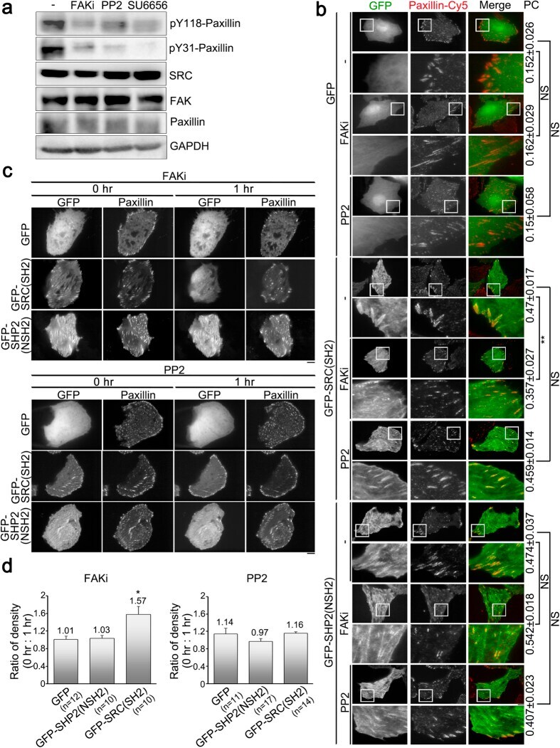
- Figure 5 Focal adhesion association of SRC controls SRC-mediated substrate phosphorylation, focal adhesion formation and cell migration. ( a ) Cell lysates from U2OS cells expressing GFP, SRC-GFP, SRCDeltaSH2-GFP, SRCY527F-GFP or SRCY527FDeltaSH2-GFP were analyzed by Western blotting for pY577-FAK, pY397-FAK, pY118-paxillin, pY31-paxillin, paxillin, GAPDH and GFP. ( b ) Cell lysates from SYF cells expressing GFP, SRC-GFP, SRCDeltaSH2-GFP, SRCY527F-GFP or SRCY527FDeltaSH2-GFP were analyzed by Western blotting for pY577-FAK, pY397-FAK, pY118-paxillin, pY31-paxillin, paxillin, GAPDH and GFP. ( c ) U2OS cells expressing GFP, SRCY527F-GFP or SRCY527FDeltaSH2-GFP (blue) were immunostained to detect F-actin (phalloidin; red) and paxillin (green), and imaged by epi-fluorescence and TIRFM, respectively. Scale bar, 10 mum. ( d ) The number of segmented paxillin-marked FAs within U2OS cells, as described in ( c ). Data are means +- s.e.m. (GFP: n = 11 cells; SRCY527F-GFP: n = 13 cells; SRCY527FDeltaSH2-GFP: n = 12 cells). ** p < 0.01; *** p < 0.001; NS, no significance. ( e ) Size distribution of segmented paxillin-marked FAs of U2OS cells, as described in ( c ). Data are means +- s.e.m. * p < 0.05; ** p < 0.01; *** p < 0.001; NS, no significance. ( f ) The migratory behavior of U2OS cells expressing GFP, SRCY527F-GFP or SRCY527FDeltaSH2-GFP for 6 hrs. Cells were plated for 16 hrs, and then monitored for 6 hrs. (right) These images delineate the trajectory of the GFP-marked cells over a

- Submitted by
- Invitrogen Antibodies (provider)
- Main image

- Experimental details
- Figure 2 Focal adhesion kinase activity, but not SRC activity, positively regulates the abundance of SRC_SH2 domain within focal adhesions. ( a ) Total cell lysate from untreated U2OS cells (-) or cells treated with FAKi (focal adhesion kinase inhibitor; 50 muM, 1 h), PP2 (SRC inhibitor; 10 muM, 1 h) or SU6656 (SRC inhibitor; 5 muM, 1 h) were analyzed by Western blotting to detect pY118-paxillin, pY31-paxillin, SRC, FAK, paxillin and GAPDH. ( b ) U2OS cells transfected with pGFP-C1, pGFP-SRC(SH2) or pGFP-SHP2(NSH2) were untreated (-) or treated with FAKi or PP2, and immunostained to detect paxillin. TIRFM images of GFP, GFP-SRC(SH2) or GFP-SHP2(NSH2) (green) and paxillin (red). Bar, 10 mum. The 20 mum x 20 mum areas indicated in the upper images are magnified in the images below. Values indicate Pearson's correlation coefficients (PC) of images of paxillin-Cy5 and GFP, GFP-SRC(SH2) or GFP-SHP2(NSH2) in U2OS cells untreated (-) or treated with FAKi or PP2. Data are means +- s.e.m. (GFP: n = 3 cells (-), n = 4 cells (FAKi), n = 6 cells (PP2); GFP-SRC(SH2): n = 7 cells (-), n = 6 cells (FAKi), n = 7 cells (PP2); GFP-SHP2(NSH2): n = 5 cells (-), n = 5 cells (FAKi), n = 4 cells (PP2)). ** p < 0.01; NS, no significance. ( c ) Time-lapse TIRFM images of U2OS cells co-transfected with mApple-paxillin (red) and pGFP-C1, pGFP-SRC(SH2) or pGFP-SHP2(NSH2) (green) during FAKi or PP2 treatment. The time is shown in hour. Bar, 10 mum. ( d ) The ratio of the average density (intensity p
- Submitted by
- Invitrogen Antibodies (provider)
- Main image

- Experimental details
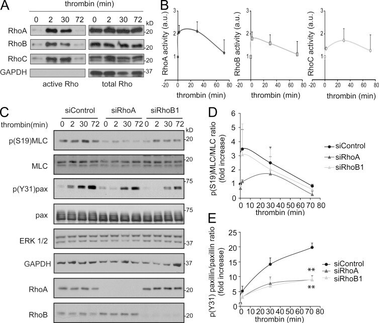
- Figure 4. Effect of RhoA and RhoB on myosin light chain and paxillin phosphorylation. (A) Pull-down assays of RhoA, RhoB, and RhoC during acute contraction induced with thrombin in HUVECs pretreated with TNF for 8 h. Graphs show active Rho normalized to total Rho levels from three different experiments. (B) TNF-pretreated, siRNA-transfected HUVECs were stimulated with thrombin for the indicated times and immunoblotted for the indicated proteins. (C and D) Quantification of phosphorylation of the indicated proteins from at least three experiments. **, P < 0.005.
- Submitted by
- Invitrogen Antibodies (provider)
- Main image

- Experimental details
- Figure 3 Phosphorylated paxillin (Ser-272) is localized in spindle poles during mitosis. ( A ) Mitotic cells were plated on coverslips and were stained with antibodies against phospho-paxillin (pS272-paxillin, pY31-paxillin or pY118-paxillin) and alpha-tublin. Nuclei were stained using DAPI. pS272-paxillin, but not pY31-paxillin and pY118-paxillin, is localized in the spindle poles. Scale bar: 10 mum. Arrows point to spindle poles. ( B ) Z stack images of cells showing connection of microtubule spindle with pS272-paxillin in the poles. Scale bar, 5 mum.
