Explore
 Validate
 Learn
Learn Western blot
Antibody data
- Antibody Data
- Antigen structure
- References [1]
- Comments [0]
- Validations
- Western blot [7]
- Immunocytochemistry [1]
- Immunoprecipitation [1]
- Immunohistochemistry [1]
Submit
Validation data
Reference
Comment
Report error
- Product number
- GTX125891 - Provider product page

- Provider
- GeneTex
- Product name
- Paxillin antibody
- Antibody type
- Polyclonal
- Reactivity
- Human, Mouse, Rat
- Host
- Rabbit
Submitted references Generation and quantitative proteomics analysis of CK2α/α'(-/-) cells.
Borgo C, Franchin C, Scalco S, Bosello-Travain V, Donella-Deana A, Arrigoni G, Salvi M, Pinna LA
Scientific reports 2017 Feb 17;7:42409
Scientific reports 2017 Feb 17;7:42409
No comments: Submit comment
Supportive validation
- Submitted by
- GeneTex (provider)
- Main image

- Experimental details
- Paxillin antibody detects PXN protein by Western blot analysis.A. 30 µg K562 whole cell lysate/extractB. 30 µg THP-1 whole cell lysate/extract7.5 % SDS-PAGEPaxillin antibody (GTX125891) dilution: 1:1000
- Validation comment
- WB
- Submitted by
- GeneTex (provider)
- Main image
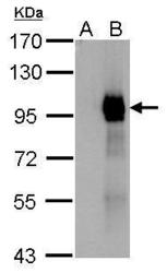
- Experimental details
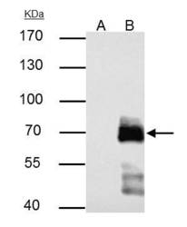
- Paxillin antibody detects Paxillin protein by western blot analysis.A. 1 ?g 293T whole cell extractB. 1 ?g whole cell extract of GFP-human Paxillin-transfected 293T cells7.5% SDS-PAGEPaxillin antibody (GTX125891) dilution: 1:10000 The HRP-conjugated anti-rabbit IgG antibody (GTX213110-01) was used to detect the primary antibody.

- Submitted by
- GeneTex (provider)
- Main image
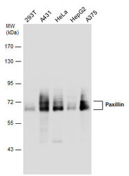
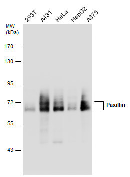
- Experimental details
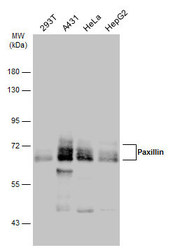
- Various whole cell extracts (30 ?g) were separated by 7.5% SDS-PAGE, and the membrane was blotted with Paxillin antibody (GTX125891) diluted at 1:5000. The HRP-conjugated anti-rabbit IgG antibody (GTX213110-01) was used to detect the primary antibody.
- Submitted by
- GeneTex (provider)
- Main image

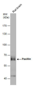
- Experimental details
- Rat tissue extract (50 ?g) was separated by 7.5% SDS-PAGE, and the membrane was blotted with Paxillin antibody (GTX125891) diluted at 1:1000. The HRP-conjugated anti-rabbit IgG antibody (GTX213110-01) was used to detect the primary antibody.
- Submitted by
- GeneTex (provider)
- Main image
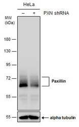
- Experimental details
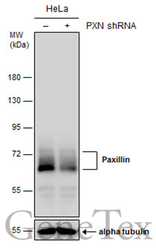
- Non-transfected (¡V) and transfected (+) HeLa whole cell extracts (30 ?g) were separated by 7.5% SDS-PAGE, and the membrane was blotted with Paxillin antibody (GTX125891) diluted at 1:6000. The HRP-conjugated anti-rabbit IgG antibody (GTX213110-01) was used to detect the primary antibody.
- Submitted by
- GeneTex (provider)
- Main image
- Experimental details
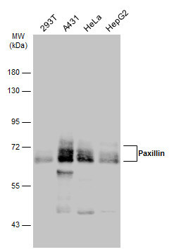
- Various whole cell extracts (30 ?g) were separated by 7.5% SDS-PAGE, and the membrane was blotted with Paxillin antibody (GTX125891) diluted at 1:5000. The HRP-conjugated anti-rabbit IgG antibody (GTX213110-01) was used to detect the primary antibody.
- Submitted by
- GeneTex (provider)
- Main image
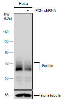
- Experimental details
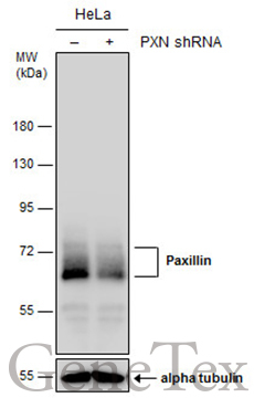
- Non-transfected (¡V) and transfected (+) HeLa whole cell extracts (30 ?g) were separated by 7.5% SDS-PAGE, and the membrane was blotted with Paxillin antibody (GTX125891) diluted at 1:6000. The HRP-conjugated anti-rabbit IgG antibody (GTX213110-01) was used to detect the primary antibody.
Supportive validation
- Submitted by
- GeneTex (provider)
- Main image

- Experimental details
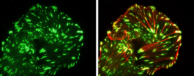
- Paxillin antibody detects Paxillin protein at cytoskeleton by immunofluorescent analysis.Sample: MDA-MB-231 cells were fixed in 4% paraformaldehyde at RT for 15 min.Green: Paxillin protein stained by Paxillin antibody (GTX125891) diluted at 1:100.Red: phalloidin staining.Courtesy of a researcher who prefers to be anonymous.
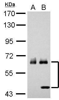
Supportive validation
- Submitted by
- GeneTex (provider)
- Main image
- Experimental details
- Paxillin antibody immunoprecipitates Paxillin protein in IP experiments.IP samples: HeLa whole cell extractA. Control with 2 £gg of preimmune Rabbit IgGB. Immunoprecipitation of Paxillin protein by 2 £gg Paxillin antibody (GTX125891)7.5 % SDS-PAGEThe immunoprecipitated Paxillin protein was detected by Paxillin antibody (GTX125891) diluted at 1:10000.[EasyBlot anti-rabbit IgG (GTX221666-01) was used as a secondary reagent]

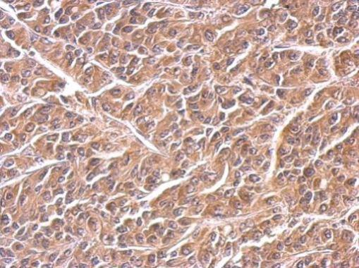
Supportive validation
- Submitted by
- GeneTex (provider)
- Main image
- Experimental details
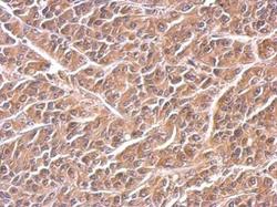
- Paxillin antibody detects PXN protein at cytosol on AGS xenograft by immunohistochemical analysis. Sample: Paraffin-embedded AGS xenograft. Paxillin antibody (GTX125891) dilution: 1:500.