Explore
 Validate
 Learn
Learn Western blot
 Immunohistochemistry
Antibody data
- Antibody Data
- Antigen structure
- References [2]
- Comments [0]
- Validations
- Western blot [1]
Submit
Validation data
Reference
Comment
Report error
- Product number
- A01940 - Provider product page

- Provider
- Boster Biological Technology
- Product name
- Anti-PKC beta 1/PRKCB Antibody Picoband™
- Antibody type
- Polyclonal
- Description
- Rabbit IgG polyclonal antibody for PKC beta 1/PRKCB detection. Tested with WB, IHC-P in Human;Mouse;Rat.
- Reactivity
- Human, Mouse, Rat
- Host
- Rabbit
- Vial size
- 100μg/vial
- Concentration
- 0.5-1mg/ml, actual concentration vary by lot. Use suggested dilution ratio to decide dilution procedure.
- Storage
- At -20°C; for one year. After reconstitution, at 4°C for one month. It can also be aliquoted and stored frozen at -20°C for a longer time. Avoid repeated freezing and thawing.
- Handling
- Add 0.2ml of distilled water will yield a concentration of 500ug/ml.
Submitted references 5-(Bis(3-(2-hydroxyethyl)-1H-indol-2-yl)methyl)-2-hydroxybenzoic acid (BHIMHA): showing a strategy of designing drug to block lung metastasis of tumors.
Apoptosis of murine melanoma B16-BL6 cells induced by quercetin targeting mitochondria, inhibiting expression of PKC-alpha and translocating PKC-delta.
Gan T, Wang Y, Zhao M, Wu J, Yang J, Peng S
Drug design, development and therapy 2016;10:711-21
Drug design, development and therapy 2016;10:711-21
Apoptosis of murine melanoma B16-BL6 cells induced by quercetin targeting mitochondria, inhibiting expression of PKC-alpha and translocating PKC-delta.
Zhang XM, Chen J, Xia YG, Xu Q
Cancer chemotherapy and pharmacology 2005 Mar;55(3):251-62
Cancer chemotherapy and pharmacology 2005 Mar;55(3):251-62
No comments: Submit comment
Supportive validation
- Submitted by
- Boster Biological Technology (provider)
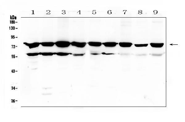
- Main image

- Experimental details
- Western blot analysis of PKC beta 1 using anti-PKC beta 1 antibody (A01940). Electrophoresis was performed on a 5-20% SDS-PAGE gel at 70V (Stacking gel) / 90V (Resolving gel) for 2-3 hours. The sample well of each lane was loaded with 50ug of sample under reducing conditions. Lane 1: rat thymus tissue lysate,Lane 2: rat spleen tissue lysate,Lane 3: rat brain tissue lysate,Lane 4: rat C6 whole cell lysate,Lane 5: mouse thymus tissue lysate,Lane 6: mouse spleen tissue lysate,Lane 7: mouse brain tissue lysate,Lane 8: mouse Neuro-2a whole cell lysate,Lane 9: mouse NIH3T3 whole cell lysate. After Electrophoresis, proteins were transferred to a Nitrocellulose membrane at 150mA for 50-90 minutes. Blocked the membrane with 5% Non-fat Milk/ TBS for 1.5 hour at RT. The membrane was incubated with rabbit anti-PKC beta 1 antigen affinity purified polyclonal antibody (Catalog # A01940) at 0.5 μg/mL overnight at 4°C, then washed with TBS-0.1%Tween 3 times with 5 minutes each and probed with a goat anti-rabbit IgG-HRP secondary antibody at a dilution of 1:10000 for 1.5 hour at RT. The signal is developed using an Enhanced Chemiluminescent detection (ECL) kit (Catalog # EK1002) with Tanon 5200 system. A specific band was detected for PKC beta 1 at approximately 77KD. The expected band size for PKC beta 1 is at 77KD.
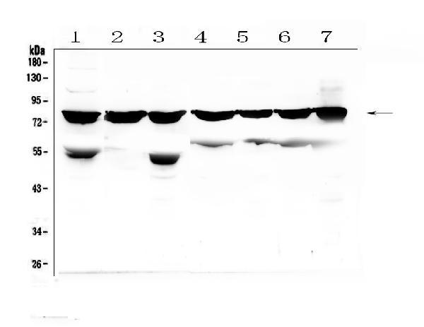
- Additional image