Explore
 Validate
 Learn
Learn Western blot
Antibody data
- Antibody Data
- Antigen structure
- References [1]
- Comments [0]
- Validations
- Western blot [1]
Submit
Validation data
Reference
Comment
Report error
- Product number
- GTX25782 - Provider product page

- Provider
- GeneTex
- Proper citation
- GeneTex Cat#GTX25782, RRID:AB_380683
- Product name
- PKC beta 1 (phospho Thr642) antibody
- Antibody type
- Polyclonal
- Reactivity
- Human, Rat
- Host
- Rabbit
Submitted references Activation of PKC isoform beta(I) at the blood-brain barrier rapidly decreases P-glycoprotein activity and enhances drug delivery to the brain.
Rigor RR, Hawkins BT, Miller DS
Journal of cerebral blood flow and metabolism : official journal of the International Society of Cerebral Blood Flow and Metabolism 2010 Jul;30(7):1373-83
Journal of cerebral blood flow and metabolism : official journal of the International Society of Cerebral Blood Flow and Metabolism 2010 Jul;30(7):1373-83
No comments: Submit comment
Supportive validation
- Submitted by
- GeneTex (provider)
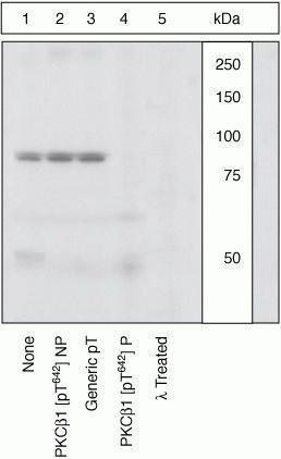
- Main image
- Experimental details
- Lysates prepared from K562 cells stimulated with PMA were resolved by SDS-PAGE on a 10% polyacrylamide gel and transferred to PVDF. Membranes were either left untreated (1-4) or treated with lambda phosphatase (5), blocked with a 5% BSA-TBST buffer for one hour at room temperature, and incubated with PKCbetaI [pT642] antibody for two hours at room temperature in a 3% BSA-TBST buffer, following prior incubation with: no peptide (1, 5), the non-phosphopeptide corresponding to the immunogen (2), a generic phosphothreonine-containing peptide (3), or, the phosphopeptide immunogen (4). After washing, membranes were incubated with goat F(ab)2 anti-rabbit IgG HRP conjugate and bands were detected. The data show that only the peptide corresponding to PKCbetaI [pT642] blocks the antibody signal. The data also show that phosphatase stripping eliminates the signal, verifying that the antibody is phospho-specific.