 Explore
 Validate
 Learn
Learn Western blot
Antibody data
- Antibody Data
- Antigen structure
- References [2]
- Comments [0]
- Validations
- Western blot [4]
- Immunocytochemistry [2]
- Other assay [2]
Submit
Validation data
Reference
Comment
Report error
- Product number
- 700286 - Provider product page

- Provider
- Invitrogen Antibodies
- Product name
- PKR Recombinant Rabbit Monoclonal Antibody (23H52L96)
- Antibody type
- Monoclonal
- Antigen
- Recombinant full-length protein
- Description
- This antibody is predicted to react with Rhesus monkey based on sequence homology.
- Antibody clone number
- 23H52L96
- Concentration
- 0.5 mg/mL
Submitted references Opposing Roles of Double-Stranded RNA Effector Pathways and Viral Defense Proteins Revealed with CRISPR-Cas9 Knockout Cell Lines and Vaccinia Virus Mutants.
Post-translational Regulation of Hexokinase Function and Protein Stability in the Aestivating Frog Xenopus laevis.
Liu R, Moss B
Journal of virology 2016 Sep 1;90(17):7864-79
Journal of virology 2016 Sep 1;90(17):7864-79
Post-translational Regulation of Hexokinase Function and Protein Stability in the Aestivating Frog Xenopus laevis.
Childers CL, Storey KB
The protein journal 2016 Feb;35(1):61-71
The protein journal 2016 Feb;35(1):61-71
No comments: Submit comment
Supportive validation
- Submitted by
- Invitrogen Antibodies (provider)
- Main image

- Experimental details
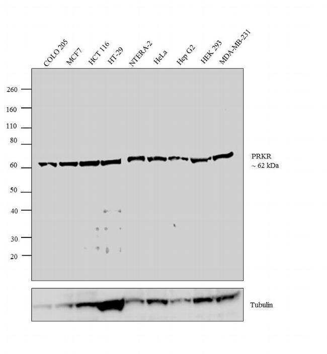
- Western blot analysis was performed on whole cell extracts (30 µg lysate) of COLO 205 (Lane 1), MCF7 (Lane 2), HCT 116 (Lane 3), HT-29 (Lane 4), NTERA-2 (Lane 5), HeLa (Lane 6), Hep G2 (Lane 7), HEK 293 (Lane 8) and MDA-MB-231 (Lane 9). The blot was probed with Anti-PRKR Rabbit Monoclonal Antibody (Product # 700286, 1-3 µg/mL) and detected by chemiluminescence using Goat anti-Rabbit IgG (H+L) Superclonal™ Secondary Antibody, HRP conjugate (Product # A27036, 0.4 µg/mL, 1:2500 dilution). A 62 kDa band corresponding to PRKR was observed across the cell lines tested. Known quantity of protein samples were electrophoresed using Novex® NuPAGE® 10 % Bis-Tris gel (Product # NP0302BOX), XCell SureLock™ Electrophoresis System (Product # EI0002) and Novex® Sharp Pre-Stained Protein Standard (Product # LC5800). Resolved proteins were then transferred onto a nitrocellulose membrane with iBlot® 2 Dry Blotting System (Product # IB21001). The membrane was probed with the relevant primary and secondary Antibody using iBind™ Flex Western Starter Kit (Product # SLF2000S). Chemiluminescent detection was performed using Pierce™ ECL Western Blotting Substrate (Product # 32106).
- Submitted by
- Invitrogen Antibodies (provider)
- Main image

- Experimental details
- Knockdown of Interferon-induced, double-stranded RNA-activated protein kinase was achieved by transfecting MCF7 with Interferon-induced, double-stranded RNA-activated protein kinase specific siRNAs (Silencer® select Product # S11187, S11185). Western Blot analysis (Fig. a) was performed using Whole cell extracts from the Interferon-induced, double-stranded RNA-activated protein kinase knockdown cells (lane 3), non-targeting scrambled siRNA transfected cells (lane 2) and untransfected cells (lane 1). The blot was probed with PKR Recombinant Rabbit Monoclonal Antibody (23H52L96) (Product # 700286, 0.5 µg/mL concentration) and Goat anti-Rabbit IgG (H+L) Superclonal™ Recombinant Secondary Antibody, HRP (Product # A27036, 1:20000 dilution). Densitometric analysis of this Western Blot is shown in histogram (Fig. b). Decrease in signal upon siRNA mediated knock down confirms that antibody is specific to Interferon-induced, double-stranded RNA-activated protein kinase.

- Submitted by
- Invitrogen Antibodies (provider)
- Main image

- Experimental details
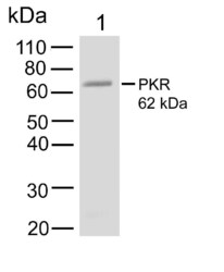
- Western Blot was performed using Anti-PKR Recombinant Rabbit Monoclonal Antibody (23H52L96) (Product # 700286) and a 62 kDa band corresponding to Interferon-induced, double-stranded RNA-activated protein kinase was observed across tested samples. Whole cell extracts (40 µg lysate) of MCF7 (Lane 1), A-431 (Lane 2), A549 (Lane 3), U-87 MG (Lane 4), NTERA-2 cl.D1 (Lane 5), Daudi (Lane 6) were electrophoresed using NuPAGE™ 4-12% Bis-Tris Protein Gel (Product # NP0321BOX). Resolved proteins were then transferred onto a nitrocellulose membrane (Product # IB23001) by iBlot® 2 Dry Blotting System (Product # IB21001). The blot was probed with the primary antibody (0.5 µg/mL concentration) and detected by chemiluminescence with Goat anti-Rabbit IgG (H+L) Superclonal™ Recombinant Secondary Antibody, HRP (Product # A27036, 1:20000 dilution) using the iBright FL 1000 (Product # A32752). Chemiluminescent detection was performed using SuperSignal™ West Dura Extended Duration Substrate (Product # 34076).

- Submitted by
- Invitrogen Antibodies (provider)
- Main image
- Experimental details
- Western blot analysis of PRKR in MCF-7 cell lysate using a PRKR recombinant rabbit monoclonal antibody (Product # 700286) at a dilution of 1 µg/mL. NBT/BCIP was used as the substrate (Product # WB7105).

Supportive validation
- Submitted by
- Invitrogen Antibodies (provider)
- Main image

- Experimental details
- Immunofluorescence analysis of Interferon-induced, double-stranded RNA-activated protein kinase was performed using 70% confluent log phase HeLa cells. The cells were fixed with 4% paraformaldehyde for 5 minutes, permeabilized with 0.1% Triton™ X-100 for 10 minutes, and blocked with 2% BSA for overnight at room temperature. The cells were labeled with PKR Recombinant Rabbit Monoclonal Antibody (23H52L96) (Product # 700286) at 5 µg/mL concentration in 0.1% BSA, incubated at 4 degree celsius overnight and then labeled with Donkey anti-Rabbit IgG (H+L) Highly Cross-Adsorbed Secondary Antibody, Alexa Fluor Plus 488 (Product # A32790), (1:2000 dilution), for 45 minutes at room temperature (Panel a: Green). Nuclei (Panel b: Blue) were stained with ProLong™ Diamond Antifade Mountant with DAPI (Product # P36962). F-actin (Panel c: Red) was stained with Rhodamine Phalloidin (Product # R415, 1:300). Panel d represents the merged image showing cytoplasm and nucleus localization. Panel e represents control cells with no primary antibody to assess background. The images were captured at 60X magnification.

- Submitted by
- Invitrogen Antibodies (provider)
- Main image

- Experimental details
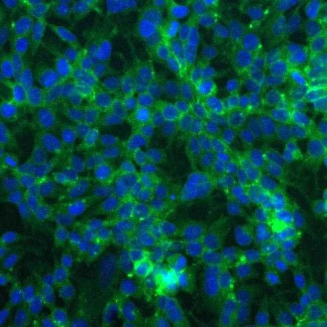
- Immunofluorescent analysis of PRKR in HEK293 cells using a PRKR recombinant rabbit monoclonal antibody (Product # 700286) at a dilution of 5 µg/mL followed by detection using an Alexa Fluor 488-conjugated goat anti-rabbit secondary antibody at a dilution of 1:1000. Cells were fixed using 4% paraformaldehyde. Cytoplasmic localization of PKR specific signal is shown in green, while nuclei were stained using SlowFade GOLD with DAPI (Product # S36938) shown in blue.
Supportive validation
- Submitted by
- Invitrogen Antibodies (provider)
- Main image

- Experimental details
- NULL

- Submitted by
- Invitrogen Antibodies (provider)
- Main image
- Experimental details
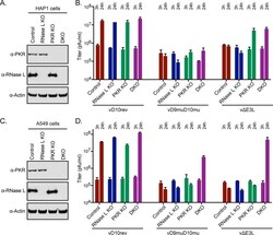
- Replication of decapping enzyme mutant and E3 deletion virus in HAP1 and A549 KO cells. (A) Absence of RNase L and PKR proteins in HAP1 KO cells. Cell lysates from HAP1 control, RNase L KO, PKR KO, and DKO cells were analyzed by Western blotting using mouse polyclonal antibody to RNase L and rabbit MAb to PKR. Antibody to actin was used as a loading control. (B) One-step virus replication. HAP1 control, RNase L KO, PKR KO, and DKO cell monolayers in 12-well plates were infected in triplicate with 5 PFU/cell of purified vD10rev, vD9muD10mu, or v(delta)E3L and harvested at 3 and 24 h. Virus titers were determined by plaque assay in BHK-21 cells. The 3-h titers represent input virus. (C and D) Procedures were the same as those described for panels A and B except that A549 control and KO cells were used. Each bar represents the standard deviation determined from three replicate infections.
