Explore
 Validate
 Learn
Learn Western blot
 Immunoprecipitation
Antibody data
- Antibody Data
- Antigen structure
- References [4]
- Comments [0]
- Validations
- Western blot [2]
- Immunocytochemistry [1]
Submit
Validation data
Reference
Comment
Report error
- Product number
- MAB1980 - Provider product page

- Provider
- R&D Systems
- Product name
- Human PKR Antibody
- Antibody type
- Monoclonal
- Description
- Protein A or G purified from hybridoma culture supernatant. Detects human PKR in Western blots.
- Reactivity
- Human
- Host
- Mouse
- Conjugate
- Unconjugated
- Isotype
- IgG
- Antibody clone number
- HL71/10
- Vial size
- 100 ug
- Concentration
- LYOPH
- Storage
- Use a manual defrost freezer and avoid repeated freeze-thaw cycles. 12 months from date of receipt, -20 to -70 °C as supplied. 1 month, 2 to 8 °C under sterile conditions after reconstitution. 6 months, -20 to -70 °C under sterile conditions after reconstitution.
Submitted references The Noncoding RNA nc886 Regulates PKR Signaling and Cytokine Production in Human Cells.
Stress-induced TRBP phosphorylation enhances its interaction with PKR to regulate cellular survival.
Altered activation of protein kinase PKR and enhanced apoptosis in dystonia cells carrying a mutation in PKR activator protein PACT.
Essential role of PACT-mediated PKR activation in tunicamycin-induced apoptosis.
Golec E, Lind L, Qayyum M, Blom AM, King BC
Journal of immunology (Baltimore, Md. : 1950) 2019 Jan 1;202(1):131-141
Journal of immunology (Baltimore, Md. : 1950) 2019 Jan 1;202(1):131-141
Stress-induced TRBP phosphorylation enhances its interaction with PKR to regulate cellular survival.
Chukwurah E, Patel RC
Scientific reports 2018 Jan 18;8(1):1020
Scientific reports 2018 Jan 18;8(1):1020
Altered activation of protein kinase PKR and enhanced apoptosis in dystonia cells carrying a mutation in PKR activator protein PACT.
Vaughn LS, Bragg DC, Sharma N, Camargos S, Cardoso F, Patel RC
The Journal of biological chemistry 2015 Sep 11;290(37):22543-57
The Journal of biological chemistry 2015 Sep 11;290(37):22543-57
Essential role of PACT-mediated PKR activation in tunicamycin-induced apoptosis.
Singh M, Fowlkes V, Handy I, Patel CV, Patel RC
Journal of molecular biology 2009 Jan 16;385(2):457-68
Journal of molecular biology 2009 Jan 16;385(2):457-68
No comments: Submit comment
Supportive validation
- Submitted by
- R&D Systems (provider)
- Main image
- Experimental details
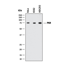
- Detection of Human PKR by Western Blot. Western blot shows lysates of HeLa human cervical epithelial carcinoma cell line, K562 human chronic myelogenous leukemia cell line, and HEK293 human embryonic kidney cell line. PVDF membrane was probed with 1 µg/mL of Mouse Anti-Human PKR Monoclonal Antibody (Catalog # MAB1980) followed by HRP-conjugated Anti-Mouse IgG Secondary Antibody (Catalog # HAF018). A specific band was detected for PKR at approximately 74 kDa (as indicated). This experiment was conducted under reducing conditions and using Immunoblot Buffer Group 1.
- Submitted by
- R&D Systems (provider)
- Main image

- Experimental details
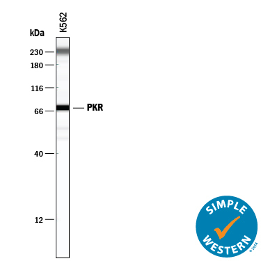
- Detection of Human PKR by Simple WesternTM. Simple Western lane view shows lysates of K562 human chronic myelogenous leukemia cell line, loaded at 0.2 mg/mL. A specific band was detected for PKR at approximately 73 kDa (as indicated) using 10 µg/mL of Mouse Anti-Human PKR Monoclonal Antibody (Catalog # MAB1980). This experiment was conducted under reducing conditions and using the 12-230 kDa separation system. Non-specific interaction with the 230 kDa Simple Western standard may be seen with this antibody.
Supportive validation
- Submitted by
- R&D Systems (provider)
- Main image
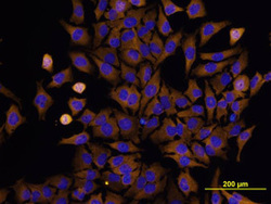
- Experimental details
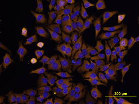
- PKR in HeLa Human Cell Line. PKR was detected in immersion fixed HeLa human cervical epithelial carcinoma cell line stimulated with rhIFN-alpha (Catalog # 11110-1) using Mouse Anti-Human PKR Monoclonal Antibody (Catalog # MAB1980) at 10 µg/mL for 3 hours at room temperature. Cells were stained using the NorthernLights™ 557-conjugated Anti-Mouse IgG Secondary Antibody (yellow; Catalog # NL007) and counterstained with DAPI (blue). View our protocol for Fluorescent ICC Staining of Cells on Coverslips.