 Explore
 Validate
 Learn
Learn Western blot
 Immunocytochemistry
Antibody data
- Antibody Data
- Antigen structure
- References [0]
- Comments [0]
- Validations
- Immunocytochemistry [2]
- Immunoprecipitation [1]
- Immunohistochemistry [5]
- Other assay [1]
Submit
Validation data
Reference
Comment
Report error
- Product number
- PA5-56292 - Provider product page

- Provider
- Invitrogen Antibodies
- Product name
- Plectin Polyclonal Antibody
- Antibody type
- Polyclonal
- Antigen
- Recombinant protein fragment
- Description
- Immunogen sequence: TEIIRQQGLA SYDYVRRRLT AEDLFEARII SLETYNLLRE GTRSLREALE AESAWCYLYG TGSVAGVYLP GSRQTLSIYQ ALKKGLLSAE VARLLLEAQA A Highest antigen sequence identity to the following orthologs: Mouse - 90%, Rat - 92%.
- Reactivity
- Human
- Host
- Rabbit
- Isotype
- IgG
- Vial size
- 100 μL
- Concentration
- 0.3 mg/mL
- Storage
- Store at 4°C short term. For long term storage, store at -20°C, avoiding freeze/thaw cycles.
No comments: Submit comment
Supportive validation
- Submitted by
- Invitrogen Antibodies (provider)
- Main image

- Experimental details
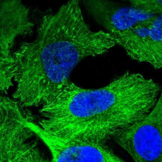
- Immunofluorescent staining of Plectin in human cell line U-251 MG shows positivity in cytoplasm, intermediate filaments & focal adhesion sites. Samples were probed using a Plectin Polyclonal Antibody (Product # PA5-56292).
- Submitted by
- Invitrogen Antibodies (provider)
- Main image

- Experimental details
- Immunofluorecent analysis of Plectin in human cell line U-251 MG using Plectin Polyclonal Antibody (Product # PA5-56292). Staining shows localization to cytosol, intermediate filaments and focal adhesion sites.

Supportive validation
- Submitted by
- Invitrogen Antibodies (provider)
- Main image
- Experimental details
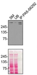
- Immunoprecipitation of Plectin was performed on U2OS cell lysates. Antibody-bead conjugates were prepared by adding 1 µg of PLEC polyclonal antibody (Product # PA5-56292) with 30 µL of protein A -Sepharose beads and rocked overnight at 4°C. 1 µg of PLEC KO lysate was incubated with antibody-bead conjugate for 2 hrs at 4°C. After multiple washes, 10% starting material (SM), 10% unbound fraction (UB) and immunoprecipitated fraction (IP) were processed for immunoblot using a PLEC monoclonal antibody. Ponceau stained transfer of blot is shown. Data courtesy of YCharOS Inc., an open science company with the mission of characterizing commercially available antibodies using knockout validation.
Supportive validation
- Submitted by
- Invitrogen Antibodies (provider)
- Main image
- Experimental details
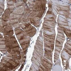
- Immunohistochemical staining of Plectin in human skeletal muscle using Plectin Polyclonal Antibody (Product # PA5-56292) shows high expression.

- Submitted by
- Invitrogen Antibodies (provider)
- Main image

- Experimental details
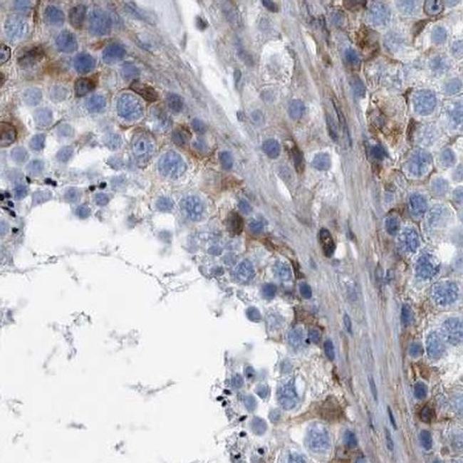
- Immunohistochemical staining of Plectin in human testis using Plectin Polyclonal Antibody (Product # PA5-56292).
- Submitted by
- Invitrogen Antibodies (provider)
- Main image

- Experimental details
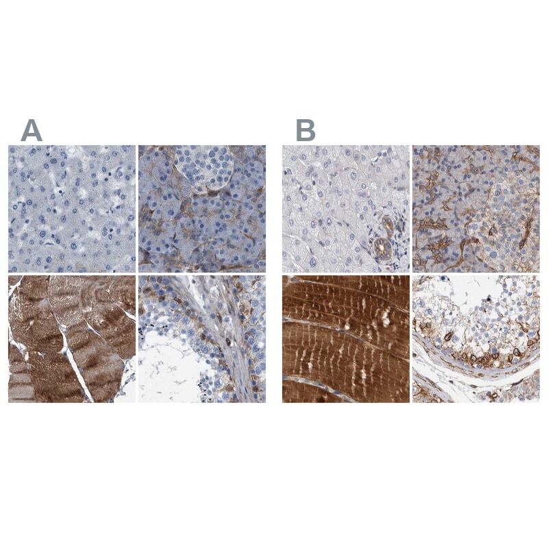
- Immunohistochemical staining of Plectin in human liver, pancreas, skeletal muscle and testis using Plectin Polyclonal Antibody (Product # PA5-56292) (A) shows similar protein distribution across tissues to an independent Plectin Polyclonal Antibody (B).
- Submitted by
- Invitrogen Antibodies (provider)
- Main image
- Experimental details
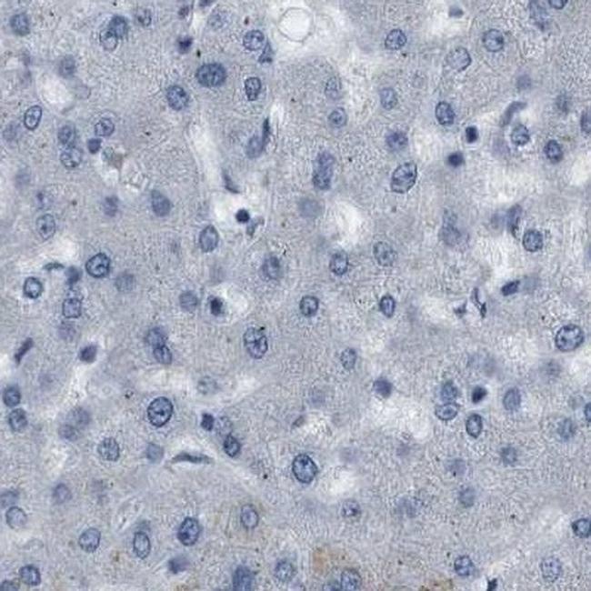
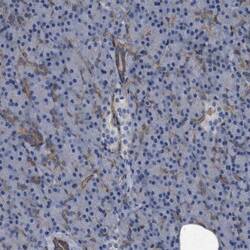
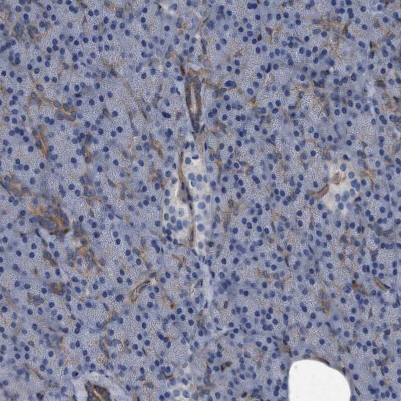
- Immunohistochemical staining of Plectin in human liver using Plectin Polyclonal Antibody (Product # PA5-56292).
- Submitted by
- Invitrogen Antibodies (provider)
- Main image
- Experimental details
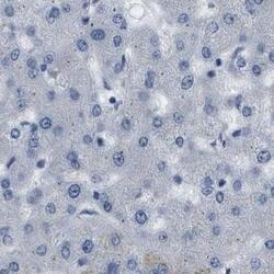
- Immunohistochemical staining of Plectin in human pancreas using Plectin Polyclonal Antibody (Product # PA5-56292) shows low expression as expected.
Supportive validation
- Submitted by
- Invitrogen Antibodies (provider)
- Main image

- Experimental details
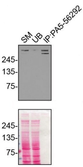
- Immunoprecipitation of Plectin was performed on U2OS cell lysates. Antibody-bead conjugates were prepared by adding 1 µg of PLEC polyclonal antibody (Product # PA5-56292) with 30 µL of protein A -Sepharose beads and rocked overnight at 4°C. 1 µg of PLEC KO lysate was incubated with antibody-bead conjugate for 2 hrs at 4°C. After multiple washes, 10% starting material (SM), 10% unbound fraction (UB) and immunoprecipitated fraction (IP) were processed for immunoblot using a PLEC monoclonal antibody. Ponceau stained transfer of blot is shown. Data courtesy of YCharOS Inc., an open science company with the mission of characterizing commercially available antibodies using knockout validation.
