Explore
 Validate
 Learn
Learn Western blot
 Immunocytochemistry
Antibody data
- Antibody Data
- Antigen structure
- References [2]
- Comments [0]
- Validations
- Western blot [4]
- Immunocytochemistry [1]
- Immunoprecipitation [1]
- Immunohistochemistry [1]
Submit
Validation data
Reference
Comment
Report error
- Product number
- GTX106334 - Provider product page

- Provider
- GeneTex
- Proper citation
- GeneTex Cat#GTX106334, RRID:AB_1241211
- Product name
- PP2A alpha antibody
- Antibody type
- Polyclonal
- Reactivity
- Human, Mouse, Rat
- Host
- Rabbit
Submitted references CME-1, a novel polysaccharide, suppresses iNOS expression in lipopolysaccharide-stimulated macrophages through ceramide-initiated protein phosphatase 2A activation.
PMC, a potent hydrophilic α-tocopherol derivative, inhibits NF-κB activation via PP2A but not IκBα-dependent signals in vascular smooth muscle cells.
Sheu JR, Chen ZC, Hsu MJ, Wang SH, Jung KW, Wu WF, Pan SH, Teng RD, Yang CH, Hsieh CY
Journal of cellular and molecular medicine 2018 Feb;22(2):999-1013
Journal of cellular and molecular medicine 2018 Feb;22(2):999-1013
PMC, a potent hydrophilic α-tocopherol derivative, inhibits NF-κB activation via PP2A but not IκBα-dependent signals in vascular smooth muscle cells.
Hsieh CY, Hsiao G, Hsu MJ, Wang YH, Sheu JR
Journal of cellular and molecular medicine 2014 Jul;18(7):1278-89
Journal of cellular and molecular medicine 2014 Jul;18(7):1278-89
No comments: Submit comment
Enhanced validation
Supportive validation
- Submitted by
- GeneTex (provider)
- Enhanced method
- Genetic validation
- Main image
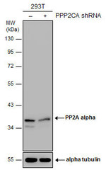
- Experimental details
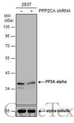
- Non-transfected (¡V) and transfected (+) 293T whole cell extracts (30 ?g) were separated by 10% SDS-PAGE, and the membrane was blotted with PP2A alpha antibody (GTX106334) diluted at 1:2000. The HRP-conjugated anti-rabbit IgG antibody (GTX213110-01) was used to detect the primary antibody.
Supportive validation
- Submitted by
- GeneTex (provider)
- Main image

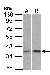
- Experimental details
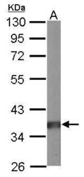
- Sample (30 ?g of whole cell lysate) A: 293T B: Molt-4 (GTX27912) 10% SDS PAGE GTX106334 diluted at 1:1500 The HRP-conjugated anti-rabbit IgG antibody (GTX213110-01) was used to detect the primary antibody.
- Submitted by
- GeneTex (provider)
- Main image
- Experimental details
- Sample (50 ?g of whole cell lysate) A: mouse brain 10% SDS PAGE GTX106334 diluted at 1:10000 The HRP-conjugated anti-rabbit IgG antibody (GTX213110-01) was used to detect the primary antibody.

- Submitted by
- GeneTex (provider)
- Main image
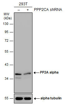
- Experimental details
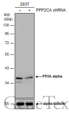
- Non-transfected (¡V) and transfected (+) 293T whole cell extracts (30 ?g) were separated by 10% SDS-PAGE, and the membrane was blotted with PP2A alpha antibody (GTX106334) diluted at 1:2000. The HRP-conjugated anti-rabbit IgG antibody (GTX213110-01) was used to detect the primary antibody.
Supportive validation
- Submitted by
- GeneTex (provider)
- Main image
- Experimental details
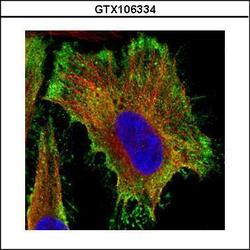
- Confocal immunofluorescence analysis (Olympus FV10i) of paraformaldehyde-fixed HeLa, using PP2A alpha(GTX106334) antibody (Green) at 1:500 dilution. Alpha-tubulin filaments were labeled with GTX11304 (Red) at 1:2000.

Supportive validation
- Submitted by
- GeneTex (provider)
- Main image
- Experimental details
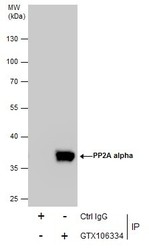
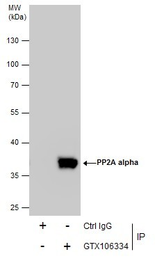
- Immunoprecipitation of PP2A alpha protein from 293T whole cell extracts using 5 £gg of PP2A alpha antibody (GTX106334).Western blot analysis was performed using PP2A alpha antibody (GTX106334).EasyBlot anti-Rabbit IgG (GTX221666-01) was used as a secondary reagent.
Supportive validation
- Submitted by
- GeneTex (provider)
- Main image
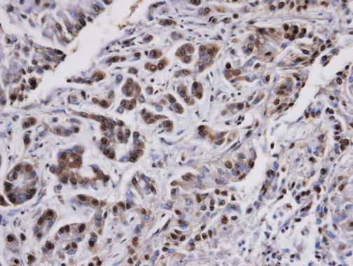
- Experimental details
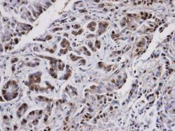
- Immunohistochemical analysis of paraffin-embedded A549 xenograft, using PPP2CA (GTX106334) antibody at 1:100 dilution.