Explore
 Validate
 Learn
Learn Western blot
Antibody data
- Antibody Data
- Antigen structure
- References [6]
- Comments [0]
- Validations
- Western blot [3]
- Other assay [2]
Submit
Validation data
Reference
Comment
Report error
- Product number
- PA5-36874 - Provider product page

- Provider
- Invitrogen Antibodies
- Product name
- Phospho-PP2A alpha (Tyr307) Polyclonal Antibody
- Antibody type
- Polyclonal
- Antigen
- Synthetic peptide
- Description
- This antibody detects endogenous protein at a molecular weight of 38 kDa. Purity is >95% by SDS-PAGE.
- Reactivity
- Human, Mouse, Rat
- Host
- Rabbit
- Isotype
- IgG
- Vial size
- 100 µL
- Concentration
- 1 mg/mL
- Storage
- Store at 4°C short term. For long term storage, store at -20°C, avoiding freeze/thaw cycles.
Submitted references Prolyl oligopeptidase inhibition reduces alpha-synuclein aggregation in a cellular model of multiple system atrophy.
The Protective Role of Prolyl Oligopeptidase (POP) Inhibition in Kidney Injury Induced by Renal Ischemia-Reperfusion.
Prolyl oligopeptidase inhibition reduces oxidative stress via reducing NADPH oxidase activity by activating protein phosphatase 2A.
PP2A(C) Phospho-Tyr(307) Antibodies Are Not Specific for this Modification but Are Sensitive to Other PP2A(C) Modifications Including Leu(309) Methylation.
Prolyl oligopeptidase inhibition activates autophagy via protein phosphatase 2A.
Salicylic Acid Targets Protein Phosphatase 2A to Attenuate Growth in Plants.
Cui H, Kilpeläinen T, Zouzoula L, Auno S, Trontti K, Kurvonen S, Norrbacka S, Hovatta I, Jensen PH, Myöhänen TT
Journal of cellular and molecular medicine 2021 Oct;25(20):9634-9646
Journal of cellular and molecular medicine 2021 Oct;25(20):9634-9646
The Protective Role of Prolyl Oligopeptidase (POP) Inhibition in Kidney Injury Induced by Renal Ischemia-Reperfusion.
Casili G, Ardizzone A, Basilotta R, Lanza M, Filippone A, Paterniti I, Esposito E, Campolo M
International journal of molecular sciences 2021 Nov 2;22(21)
International journal of molecular sciences 2021 Nov 2;22(21)
Prolyl oligopeptidase inhibition reduces oxidative stress via reducing NADPH oxidase activity by activating protein phosphatase 2A.
Eteläinen T, Kulmala V, Svarcbahs R, Jäntti M, Myöhänen TT
Free radical biology & medicine 2021 Jun;169:14-23
Free radical biology & medicine 2021 Jun;169:14-23
PP2A(C) Phospho-Tyr(307) Antibodies Are Not Specific for this Modification but Are Sensitive to Other PP2A(C) Modifications Including Leu(309) Methylation.
Frohner IE, Mudrak I, Schüchner S, Anrather D, Hartl M, Sontag JM, Sontag E, Wadzinski BE, Preglej T, Ellmeier W, Ogris E
Cell reports 2020 Mar 3;30(9):3171-3182.e6
Cell reports 2020 Mar 3;30(9):3171-3182.e6
Prolyl oligopeptidase inhibition activates autophagy via protein phosphatase 2A.
Svarcbahs R, Jäntti M, Kilpeläinen T, Julku UH, Urvas L, Kivioja S, Norrbacka S, Myöhänen TT
Pharmacological research 2020 Jan;151:104558
Pharmacological research 2020 Jan;151:104558
Salicylic Acid Targets Protein Phosphatase 2A to Attenuate Growth in Plants.
Tan S, Abas M, Verstraeten I, Glanc M, Molnár G, Hajný J, Lasák P, Petřík I, Russinova E, Petrášek J, Novák O, Pospíšil J, Friml J
Current biology : CB 2020 Feb 3;30(3):381-395.e8
Current biology : CB 2020 Feb 3;30(3):381-395.e8
No comments: Submit comment
Supportive validation
- Submitted by
- Invitrogen Antibodies (provider)
- Main image

- Experimental details
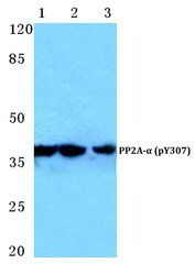
- Western blot analysis of Phospho-PP2A alpha pTyr307 using Phospho-PP2A alpha pTyr307 polyclonal antibody (Product # PA5-36874) at a dilution of 1:500. Lane 1: HEK293T cells treated with EGF (0.1 ng/mL, 30 min), Lane 2: Mouse brain tissue lysate, Lane 3: H9C2 cells treated with EGF (0.1 ng/mL, 30 min).

- Submitted by
- Invitrogen Antibodies (provider)
- Main image
- Experimental details
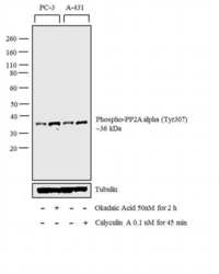
- Western blot analysis was performed on whole cell extracts (30 µg lysate) of PC-3 (Lane 1), PC-3 treated with Okadaic Acid (50nM for 2 h) (Lane 2), A-431 (Lane 3) and A-431 treated with Calyculin A (0.1 uM for 45 min) (Lane 4). The blot was probed with Anti-Phospho-PP2A alpha (Tyr307) Polyclonal Antibody (Product # PA5-36874, 1:1000 dilution) and detected by chemiluminescence using Goat anti-Rabbit IgG (H+L) Superclonal™ Secondary Antibody, HRP conjugate (Product # A27036, 0.25 µg/mL, 1:4000 dilution). A 36 kDa band corresponding to Phospho-PP2A alpha (Tyr307) was observed across the cell lines tested and enhanced upon treatment.

- Submitted by
- Invitrogen Antibodies (provider)
- Main image
- Experimental details
- Western blot analysis of Phospho-PP2A alpha (Tyr307) in Lane 1: HEK293T cell treated with EGF (0.1 ng/mL, 30 min), Lane 2: mouse brain tissue lysate, Lane 3: H9C2 cell treated with EGF (0.1 ng/mL, 30 min). Samples were incubated with Phospho-PP2A alpha (Tyr307) polyclonal antibody (Product # PA5-36874) at a dilution of 1:500.

Supportive validation
- Submitted by
- Invitrogen Antibodies (provider)
- Main image

- Experimental details
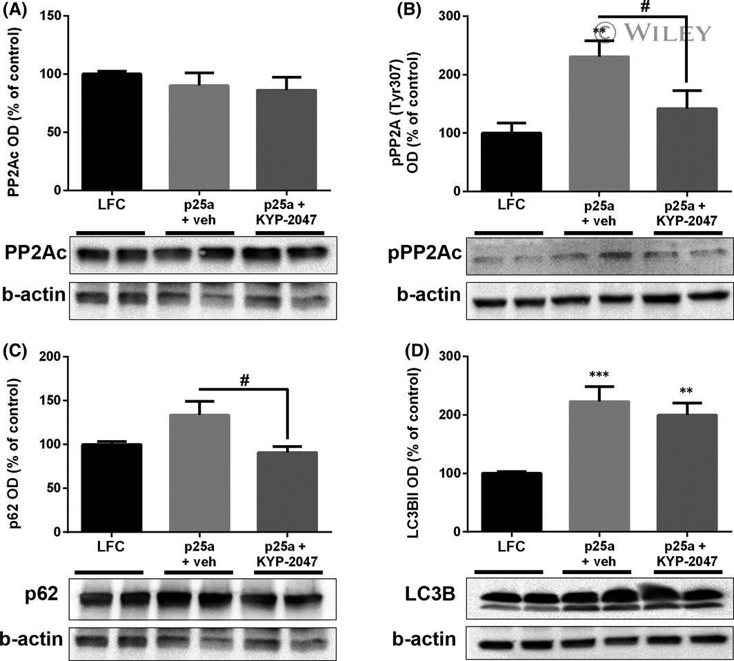
- 4 FIGURE PREP inhibition reduces PP2A phosphorylation and activates autophagy in p25alpha transfected OLN-AS7 cells. p25alpha transfection did not alter the levels of catalytic subunit of PP2A (A; PP2Ac) but Tyr307 phosphorylated PP2Ac (B; pPP2Ac) was significantly elevated after transfection and vehicle treatment (B). 10 uM KYP-2047 significantly reduced pPP2Ac levels compared to p25alpha+vehicle but did not change the levels of PP2Ac (A-B). The protein accumulation marker, p62, was significantly decreased in p25alpha transfected and KYP-2047 treated cells compared to vehicle treatment (C). Autophagosome marker LC3BII was elevated in p25alpha transfected cells and remained elevated after 10 uM KYP-2047 treatment (D). Data are presented as mean +- SEM. *, p < 0.05; **, p < 0.01; ***, p < 0.001, 1-way ANOVA with Tukey's post hoc test (compared to vehicle). #, p < 0.05; 1-way ANOVA with Tukey's post hoc test (p25alpha+veh vs. p25alpha+KYP-2047). See figure S5for uncut blots. Representative blots in Figure 4A and C are from the same membrane and therefore share the beta-actin loading control
- Submitted by
- Invitrogen Antibodies (provider)
- Main image
- Experimental details
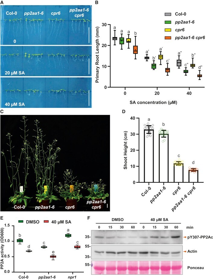
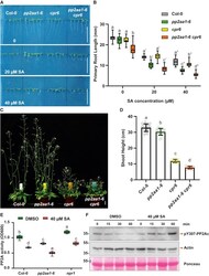
- Figure 5 Genetic Analysis of pp2aa1-6 and cpr6 Mutations, and SA Inhibits PP2A Activity In Planta (A) Representative images showing the enhanced sensitivity of pp2aa1-6 to SA. Col-0, pp2aa1-6 , cpr6 , and pp2aa1-6 cpr6 seedlings were grown on plates with different concentrations of SA for 7 days. Scale bars, 2 cm. (B) The root growth analysis revealed that the cpr6 mutation decreased the primary root length and increased the SA sensitivity of pp2aa1-6 . n = 16. Different letters represent significant difference; p < 0.05; by one-way ANOVA with a Tukey multiple comparison test. (C and D) The pp2aa1-6 mutation enhances the stunted shoot phenotype of cpr6 . Col-0, pp2aa1-6 , cpr6 , and pp2aa1-6 cpr6 plants were grown for 38 days, and representative plants are shown (C). Scale bar, 2 cm. (D) The height of plants was measured and shown as dot plots. Dots represent individual values, and lines indicate mean +- SD. n = 16. Different letters represent significant difference; p < 0.05; by one-way ANOVA with a Tukey multiple comparison test. (E) SA treatment decreased the total PP2A activity in planta . Col-0, pp2aa1-6 , and npr1 seedlings were grown on plates containing DMSO or 40 muM SA for 5 days and then sampled for protein isolation and PP2A activity measurement. n = 6. Different letters represent significant difference; p < 0.05; by one-way ANOVA with a Tukey multiple comparison test. (F) SA treatment increased the phosphorylation of the PP2A catalytic subunits (PP2Ac), suggestin