Explore
 Validate
 Learn
Learn Western blot
 Immunocytochemistry
Antibody data
- Antibody Data
- Antigen structure
- References [1]
- Comments [0]
- Validations
- Western blot [1]
Submit
Validation data
Reference
Comment
Report error
- Product number
- M01893-1 - Provider product page

- Provider
- Boster Biological Technology
- Product name
- Anti-PP2A-alpha/PPP2CA Antibody Picoband™ (monoclonal, 3B6)
- Antibody type
- Monoclonal
- Description
- Mouse IgG monoclonal antibody for PP2A-alpha/PPP2CA detection. Tested with WB, IHC-P, ICC/IF, FCM in Human;Monkey;Mouse;Rat.
- Reactivity
- Human, Mouse, Rat, Simian
- Host
- Mouse
- Isotype
- IgG
- Antibody clone number
- 3B6
- Vial size
- 100μg/vial
- Concentration
- Add 0.2ml of distilled water will yield a concentration of 500ug/ml.
- Storage
- At -20°C for one year. After reconstitution, at 4°C for one month. It can also be aliquoted and stored frozen at -20°C for a longer time. Avoid repeated freezing and thawing.
- Handling
- Add 0.2ml of distilled water will yield a concentration of 500ug/ml.
Submitted references Role of NMDA receptors in noise-induced tau hyperphosphorylation in rat hippocampus and prefrontal cortex.
Li K, Jia H, She X, Cui B, Zhang N, Chen X, Xu C, An G, Ma Q
Journal of the neurological sciences 2014 May 15;340(1-2):191-7
Journal of the neurological sciences 2014 May 15;340(1-2):191-7
No comments: Submit comment
Supportive validation
- Submitted by
- Boster Biological Technology (provider)
- Main image
- Experimental details
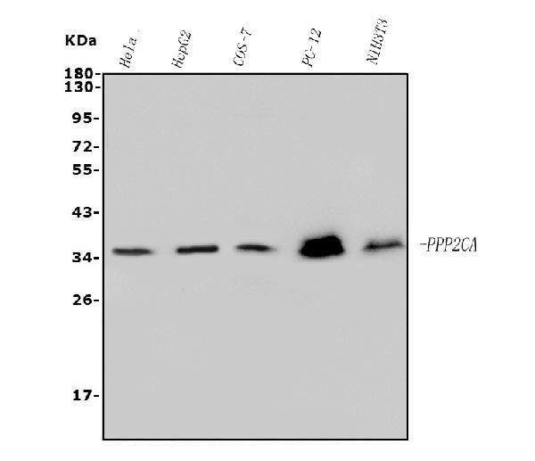
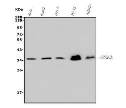
- Western blot analysis of PP2A-alpha/PPP2CA using anti-PP2A-alpha/PPP2CA antibody (M01893-1). Electrophoresis was performed on a 5-20% SDS-PAGE gel at 70V (Stacking gel) / 90V (Resolving gel) for 2-3 hours. The sample well of each lane was loaded with 50ug of sample under reducing conditions. Lane 1: human HELA whole cell lysates, Lane 2: human HEPG2 whole cell lysates, Lane 3: monkey COS-7 whole cell lysates, Lane 4: rat PC-12 whole cell lysates, Lane 5: mouse NIH/3T3 whole cell lysates. After Electrophoresis, proteins were transferred to a Nitrocellulose membrane at 150mA for 50-90 minutes. Blocked the membrane with 5% Non-fat Milk/ TBS for 1.5 hour at RT. The membrane was incubated with mouse anti- PP2A-alpha/PPP2CA antigen affinity purified monoclonal antibody (Catalog # M01893-1) at 0.5 μg/mL overnight at 4°C, then washed with TBS-0.1%Tween 3 times with 5 minutes each and probed with a goat anti-mouse IgG-HRP secondary antibody at a dilution of 1:10000 for 1.5 hour at RT. The signal is developed using an Enhanced Chemiluminescent detection (ECL) kit (Catalog # EK1001) with Tanon 5200 system. A specific band was detected for PP2A-alpha/PPP2CA at approximately 36KD. The expected band size for PP2A-alpha/PPP2CA is at 36KD.
- Additional image