Explore
 Validate
 Learn
Learn Western blot
 Immunocytochemistry
Antibody data
- Antibody Data
- Antigen structure
- References [2]
- Comments [0]
- Validations
- Immunocytochemistry [6]
- Immunohistochemistry [1]
- Flow cytometry [4]
- Chromatin Immunoprecipitation [2]
- Other assay [2]
Submit
Validation data
Reference
Comment
Report error
- Product number
- 700183 - Provider product page

- Provider
- Invitrogen Antibodies
- Product name
- PYK2 Recombinant Rabbit Monoclonal Antibody (9H12L1)
- Antibody type
- Monoclonal
- Antigen
- Recombinant full-length protein
- Description
- This antibody is predicted to react with bovine, canine, chimpanzee, equine, mouse, orangutan, rat and Rhesus monkey based on sequence homology. Intact IgG appears on a non-reducing gel as ~150 kDa band and upon reduction generating a ~25 kDa light chain band and a ~50 kDa heavy chain. Recombinant rabbit monoclonal antibodies are produced using in vitro expression systems. The expression systems are developed by cloning in the specific antibody DNA sequences from immunoreactive rabbits. Then, individual clones are screened to select the best candidates for production. The advantages of using recombinant rabbit monoclonal antibodies include: better specificity and sensitivity, lot-to-lot consistency, animal origin-free formulations, and broader immunoreactivity to diverse targets due to larger rabbit immune repertoire.
- Reactivity
- Human
- Host
- Rabbit
- Isotype
- IgG
- Antibody clone number
- 9H12L1
- Vial size
- 100 μg
- Concentration
- 0.5 mg/mL
- Storage
- Store at 4°C short term. For long term storage, store at -20°C, avoiding freeze/thaw cycles.
Submitted references PYK2 promotes HER2-positive breast cancer invasion.
CD33 modulates TREM2: convergence of Alzheimer loci.
Al-Juboori SI, Vadakekolathu J, Idri S, Wagner S, Zafeiris D, Pearson JR, Almshayakhchi R, Caraglia M, Desiderio V, Miles AK, Boocock DJ, Ball GR, Regad T
Journal of experimental & clinical cancer research : CR 2019 May 22;38(1):210
Journal of experimental & clinical cancer research : CR 2019 May 22;38(1):210
CD33 modulates TREM2: convergence of Alzheimer loci.
Chan G, White CC, Winn PA, Cimpean M, Replogle JM, Glick LR, Cuerdon NE, Ryan KJ, Johnson KA, Schneider JA, Bennett DA, Chibnik LB, Sperling RA, Bradshaw EM, De Jager PL
Nature neuroscience 2015 Nov;18(11):1556-8
Nature neuroscience 2015 Nov;18(11):1556-8
No comments: Submit comment
Supportive validation
- Submitted by
- Invitrogen Antibodies (provider)
- Main image

- Experimental details
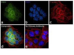
- Immunofluorescence analysis of FAK2 was performed using 70% confluent log phase A-431 cells. The cells were fixed with 4% paraformaldehyde for 10 minutes, permeabilized with 0.1% Triton™ X-100 for 10 minutes, and blocked with 1% BSA for 1 hour at room temperature. The cells were labeled with FAK2 (9H12L1) Recombinant Rabbit Monoclonal Antibody (Product # 700183) at 5 µg/mL in 0.1% BSA and incubated overnight at 4 degree and then labeled with Goat anti-Rabbit IgG (H+L) Recombinant Superclonal™ Secondary Antibody, Alexa Fluor® 488 conjugate (Product # A27034) at a dilution of 1:2000 for 45 minutes at room temperature (Panel a: green). Nuclei (Panel b: blue) were stained with SlowFade® Gold Antifade Mountant with DAPI (Product # S36938). F-actin (Panel c: red) was stained with Rhodamine Phalloidin (Product # R415, 1:300). Panel d represents the merged image showing nuclear and cytoplasmic localization. Panel e represents control cells with no primary antibody to assess background. The images were captured at 60X magnification.

- Submitted by
- Invitrogen Antibodies (provider)
- Main image

- Experimental details
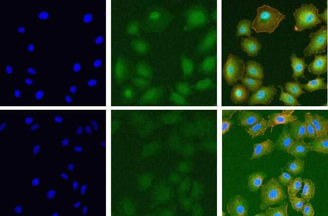
- Immunofluorescent analysis of FAK2 in A549 cells using a FAK2 recombinant rabbit monoclonal antibody (Product # 700183) at a dilution of 20 µg/mL in the absence of peptide (top) or in the presence of the immunogenic peptide (bottom) followed by detection using an Alexa Fluor 488-conjugated goat anti-rabbit secondary antibody at a dilution of 1:1000 and actin staining using Alexa Fluor 568 Phalloidin (Product # A12380). Hoechst only (left), AF488 signal only (middle) and composite image with Phalloidin (right).
- Submitted by
- Invitrogen Antibodies (provider)
- Main image
- Experimental details
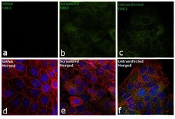
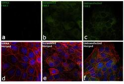
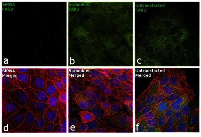
- Knockdown of FAK2 was achieved by transfecting A-431 cells with FAK2 specific siRNA (Silencer® select Product # S5007). Immunofluorescence analysis was performed on A-431cells (untransfected, panel c,f), transfected with non-specific scrambled siRNA (panels b,e) and transfected with FAK2 specific siRNA (panel a,d). Cells were fixed, permeabilized, and labelled with FAK2 Antibody (9H12L1), Recombinant Rabbit Monoclonal (Product # 700183, 5 µg/mL), followed by Goat anti-Rabbit IgG (H+L) Superclonal™ Secondary Antibody, Alexa Fluor® 488 conjugate (Product # A27034, 1:2000). Nuclei (blue) were stained using SlowFade® Gold Antifade Mountant with DAPI (Product # S36938), and Rhodamine Phalloidin (Product # R415, 1:300) was used for cytoskeletal F-actin (red) staining. Reduction of specific signal was observed upon siRNA mediated knockdown (panel a,d) confirming specificity of the antibody to FAK2 (green). The images were captured at 60X magnification.

- Submitted by
- Invitrogen Antibodies (provider)
- Main image

- Experimental details
- Immunofluorescent analysis of FAK2 in A549 cells using a FAK2 recombinant rabbit monoclonal antibody (Product # 700183) at a dilution of 20 µg/mL in the absence of peptide (top) or in the presence of the immunogenic peptide (bottom) followed by detection using an Alexa Fluor 488-conjugated goat anti-rabbit secondary antibody at a dilution of 1:1000 and actin staining using Alexa Fluor 568 Phalloidin (Product # A12380). Hoechst only (left), AF488 signal only (middle) and composite image with Phalloidin (right).

- Submitted by
- Invitrogen Antibodies (provider)
- Main image
- Experimental details
- Immunofluorescence analysis of FAK2 was performed using 70% confluent log phase A-431 cells. The cells were fixed with 4% paraformaldehyde for 10 minutes, permeabilized with 0.1% Triton™ X-100 for 10 minutes, and blocked with 1% BSA for 1 hour at room temperature. The cells were labeled with FAK2 (9H12L1) Recombinant Rabbit Monoclonal Antibody (Product # 700183) at 5 µg/mL in 0.1% BSA and incubated overnight at 4 degree and then labeled with Goat anti-Rabbit IgG (Heavy Chain) Recombinant Superclonal™ Secondary Antibody, Alexa Fluor® 488 conjugate (Product # A27034) at a dilution of 1:2000 for 45 minutes at room temperature (Panel a: green). Nuclei (Panel b: blue) were stained with SlowFade® Gold Antifade Mountant with DAPI (Product # S36938). F-actin (Panel c: red) was stained with Rhodamine Phalloidin (Product # R415, 1:300). Panel d represents the merged image showing nuclear and cytoplasmic localization. Panel e represents control cells with no primary antibody to assess background. The images were captured at 60X magnification.

- Submitted by
- Invitrogen Antibodies (provider)
- Main image
- Experimental details
- Knockdown of FAK2 was achieved by transfecting A-431 cells with FAK2 specific siRNA (Silencer® select Product # S5007). Immunofluorescence analysis was performed on A-431cells (untransfected, panel c,f), transfected with non-specific scrambled siRNA (panels b,e) and transfected with FAK2 specific siRNA (panel a,d). Cells were fixed, permeabilized, and labelled with FAK2 Antibody (9H12L1), Recombinant Rabbit Monoclonal (Product # 700183, 5 µg/mL), followed by Goat anti-Rabbit IgG (Heavy Chain) Superclonal™ Secondary Antibody, Alexa Fluor® 488 conjugate (Product # A27034, 1:2000). Nuclei (blue) were stained using SlowFade® Gold Antifade Mountant with DAPI (Product # S36938), and Rhodamine Phalloidin (Product # R415, 1:300) was used for cytoskeletal F-actin (red) staining. Reduction of specific signal was observed upon siRNA mediated knockdown (panel a,d) confirming specificity of the antibody to FAK2 (green). The images were captured at 60X magnification.
Supportive validation
- Submitted by
- Invitrogen Antibodies (provider)
- Main image
- Experimental details
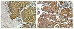
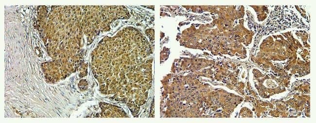
- Immunohistochemistry analysis of FAK2 in formalin-fixed, paraffin-embedded human breast (left) and lung carcimona (right) using a FAK2 monoclonal antibody (Product # 700183) at a dilution of 5 µg/mL. Tissues were pretreated with EDTA and staining was visualized using DAB. Images were taken at a magnification of 40x. Results show cytoplasmic staining in tumor cells.
Supportive validation
- Submitted by
- Invitrogen Antibodies (provider)
- Main image
- Experimental details
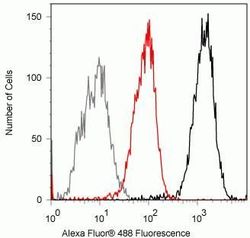
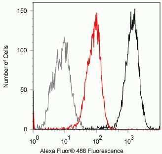
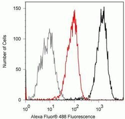
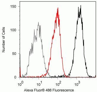
- Flow cytometry analysis of FAK2 in Jurkat cells using a FAK2 recombinant rabbit monoclonal antibody (Product # 700183) at a dilution of 0.5 µg. Cells were fixed and permeabilized using FIX & PERM (Product # GAS-004) reagent, and detection was performed using an Alexa Fluor 488 goat anti-rabbit IgG (black) compared to a control without primary antibody (gray). Pre-incubation with the immunogenic peptide decreased the signal (red).

- Submitted by
- Invitrogen Antibodies (provider)
- Main image
- Experimental details
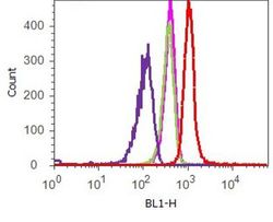
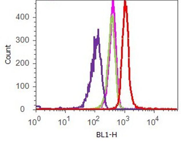
- Flow cytometry analysis of Pyk2 was done on HeLa cells. Cells were fixed with 70% ethanol for 10 minutes, permeabilized with 0.25% Tritonª X-100 for 20 minutes, and blocked with 5% BSA for 1 hour at room temperature. Cells were labeled with ABfinityª Pyk2 Recombinant Rabbit Monoclonal Antibody (700183, red histogram) or with rabbit isotype control (pink histogram) at 2 µg-4 µg/million cells in 2.5% BSA. After incubation at room temperature for 2-3 hours, the cells were labeled with Alexa Fluor¨ 488 Goat Anti-Rabbit Secondary Antibody (A11008) at a dilution of 1:400 for 30 minutes at room temperature. The representative 10,000 cells were acquired and analyzed for each sample using an Attune¨ Acoustic Focusing Cytometer. The purple histogram represents unstained control cells and the green histogram represents no-primary-antibody control.
- Submitted by
- Invitrogen Antibodies (provider)
- Main image
- Experimental details
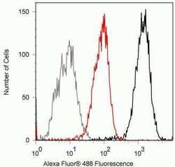
- Flow cytometry analysis of FAK2 in Jurkat cells using a FAK2 recombinant rabbit monoclonal antibody (Product # 700183) at a dilution of 0.5 µg. Cells were fixed and permeabilized using FIX & PERM (Product # GAS-004) reagent, and detection was performed using an Alexa Fluor 488 goat anti-rabbit IgG (black) compared to a control without primary antibody (gray). Pre-incubation with the immunogenic peptide decreased the signal (red).
- Submitted by
- Invitrogen Antibodies (provider)
- Main image
- Experimental details
- Flow cytometry analysis of FAK2 in Jurkat cells using a FAK2 recombinant rabbit monoclonal antibody (Product # 700183) at a dilution of 0.5 µg. Cells were fixed and permeabilized using FIX & PERM (Product # GAS-004) reagent, and detection was performed using an Alexa Fluor 488 goat anti-rabbit IgG (black) compared to a control without primary antibody (gray). Pre-incubation with the immunogenic peptide decreased the signal (red).
Supportive validation
- Submitted by
- Invitrogen Antibodies (provider)
- Main image
- Experimental details
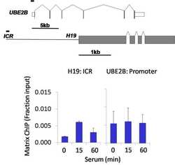
- Chromatin immunoprecipitation analysis of FAK2/PYK2 was performed using cross-linked chromatin from 1 x 10^6 HCT116 human colon carcinoma cells treated with serum for 0, 15, and 60 minutes. Immunoprecipitation was performed using a multiplex microplate Matrix ChIP assay (see reference for Matrix ChIP protocol: http://www.ncbi.nlm.nih.gov/pubmed/22098709) with 1.0 µL/100 µL well volume of a FAK2/PYK2 rabbit monoclonal antibody (Product # 700183). Chromatin aliquots from ~1 x 10^5 cells were used per ChIP pull-down. Quantitative PCR data were done in quadruplicate using 1 µL of eluted DNA in 2 µL SYBR real-time PCR reactions containing primers to amplify the promoter region of human UBE2B, or the imprinting control region (ICR) of the human H19 locus. PCR calibration curves were generated for each primer pair from a dilution series of sheared total genomic DNA. Quantitation of immunoprecipitated chromatin is presented as signal relative to the total amount of input chromatin. Results represent the mean +/- SEM for three experiments. A schematic representation of the human UBE2B and H19 loci are shown above the data where boxes represent exons (grey boxes = translated regions, white boxes = untranslated regions), the zigzag lines represent introns, and the straight line represents upstream sequence. Regions amplified by UBE2B and H19 primers are represented by black bars. Data courtesy of the Innovators Program.

- Submitted by
- Invitrogen Antibodies (provider)
- Main image

- Experimental details
- Chromatin immunoprecipitation analysis of FAK2/PYK2 was performed using cross-linked chromatin from 1 x 10^6 HCT116 human colon carcinoma cells treated with serum for 0, 15, and 60 minutes. Immunoprecipitation was performed using a multiplex microplate Matrix ChIP assay (see reference for Matrix ChIP protocol: http://www.ncbi.nlm.nih.gov/pubmed/22098709) with 1.0 µL/100 µL well volume of a FAK2/PYK2 rabbit monoclonal antibody (Product # 700183). Chromatin aliquots from ~1 x 10^5 cells were used per ChIP pull-down. Quantitative PCR data were done in quadruplicate using 1 µL of eluted DNA in 2 µL SYBR real-time PCR reactions containing primers to amplify the promoter region of human UBE2B, or the imprinting control region (ICR) of the human H19 locus. PCR calibration curves were generated for each primer pair from a dilution series of sheared total genomic DNA. Quantitation of immunoprecipitated chromatin is presented as signal relative to the total amount of input chromatin. Results represent the mean +/- SEM for three experiments. A schematic representation of the human UBE2B and H19 loci are shown above the data where boxes represent exons (grey boxes = translated regions, white boxes = untranslated regions), the zigzag lines represent introns, and the straight line represents upstream sequence. Regions amplified by UBE2B and H19 primers are represented by black bars. Data courtesy of the Innovators Program.

Supportive validation
- Submitted by
- Invitrogen Antibodies (provider)
- Main image
- Experimental details
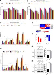
- Fig. 1 Effect of metformin on cell proliferation and apoptosis of breast cancer cell lines representing different phenotypes of breast cancer. a , b Effect of different concentrations of metformin on cell proliferation of BT-474, MCF-7, MDA-MB-231, MDA-MB-468 and SkBr3, 24 h and 48 h post-treatment. N = 3 (6 replicates). c , d Effect of different concentrations of metformin on apoptosis of BT-474, MCF-7, MDA-MB-231, MDA-MB-468 and SkBr3, 24 h and 48 h post-treatment. N = 3 (2 replicates). The statistical values are provided as Additional file 4 : Data S1. e Heatmap of microarray analysis showing upregulated (in red) and downregulated genes (in blue) in untreated vs. treated cells. Bonferroni corrected P value

- Submitted by
- Invitrogen Antibodies (provider)
- Main image

- Experimental details
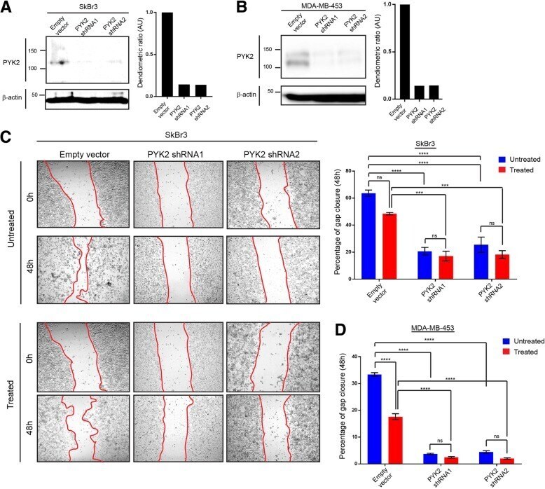
- Fig. 2 Effect of PYK2 knockdown on cell migration of the HER2+/ER-/PR- breast cancer cell lines SkBr3 and MDA-MB-453. a, b Immunoblot images representing PYK2 expression SkBr3 and MDA-MB-453 control (pLKO.1 empty vector) and PYK2 knockdown cells. N= 3 (3 replicates). c Wound healing assay (scratch assay) using SkBr3 control and PYK2 knockdown metformin treated and untreated cells, and the corresponding data quantifying gap closure at time points 0 and 48h following scratching. Anova ****P=