Explore
 Validate
 Learn
Learn Western blot
 Immunocytochemistry
Antibody data
- Antibody Data
- Antigen structure
- References [4]
- Comments [0]
- Validations
- Western blot [1]
Submit
Validation data
Reference
Comment
Report error
- Product number
- GTX13593 - Provider product page

- Provider
- GeneTex
- Proper citation
- GeneTex Cat#GTX13593, RRID:AB_372365
- Product name
- STAT5A (phospho Tyr694) antibody
- Antibody type
- Polyclonal
- Reactivity
- Human, Mouse
- Host
- Rabbit
Submitted references Phosphorylation of calcium/calmodulin-stimulated protein kinase II at T286 enhances invasion and migration of human breast cancer cells.
Cabozantinib is selectively cytotoxic in acute myeloid leukemia cells with FLT3-internal tandem duplication (FLT3-ITD).
Granulocyte colony-stimulating factor potentiates all-trans retinoic acid-induced granulocytic differentiation in acute promyelocytic leukemia cell line HT93A.
Erythropoietin protects neuroblastoma cells against etoposide and vincristine by activating ERK and AKT pathways but has no effect in kidney cells.
Chi M, Evans H, Gilchrist J, Mayhew J, Hoffman A, Pearsall EA, Jankowski H, Brzozowski JS, Skelding KA
Scientific reports 2016 Sep 8;6:33132
Scientific reports 2016 Sep 8;6:33132
Cabozantinib is selectively cytotoxic in acute myeloid leukemia cells with FLT3-internal tandem duplication (FLT3-ITD).
Lu JW, Wang AN, Liao HA, Chen CY, Hou HA, Hu CY, Tien HF, Ou DL, Lin LI
Cancer letters 2016 Jul 1;376(2):218-25
Cancer letters 2016 Jul 1;376(2):218-25
Granulocyte colony-stimulating factor potentiates all-trans retinoic acid-induced granulocytic differentiation in acute promyelocytic leukemia cell line HT93A.
Uchino Y, Iriyama N, Hatta Y, Takei M
Cancer cell international 2015;15:30
Cancer cell international 2015;15:30
Erythropoietin protects neuroblastoma cells against etoposide and vincristine by activating ERK and AKT pathways but has no effect in kidney cells.
Vazquez-Mellado MJ, Aguilar C, Rocha-Zavaleta L
Life sciences 2015 Sep 15;137:142-9
Life sciences 2015 Sep 15;137:142-9
No comments: Submit comment
Supportive validation
- Submitted by
- GeneTex (provider)
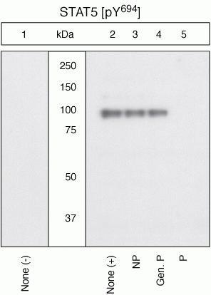
- Main image
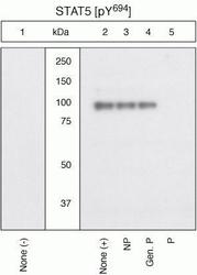
- Experimental details
- Extracts of 3T3-L1 cells untreated (1) or treated with 20 ng/mL LIF for 10 minutes (2-5) were resolved by SDS-PAGE on a 10% Tris-glycine gel and transferred to PVDF. The membrane was blocked with a 5% BSA-TBST buffer for one hour at room temperature, then incubated with the STAT5 [pY694] antibody for two hours at room temperature in a 3% BSA-TBST buffer, following prior incubation with: no peptide (1, 2), the non-phosphopeptide corresponding to the phosphopeptide immunogen (3), a generic phosphotyrosine-containing peptide (4), or the phosphopeptide immunogen (5). After washing, the membrane was incubated with goat F(ab)2 anti-rabbit IgG HRP conjugate and signals were detected. The data show that only the phosphopeptide corresponding to STAT5 [pY694] blocks the signal, verifying the specificity of the antibody. The data also show the up-regulation of the signal by treatment with LIF in this cell system.