Explore
 Validate
 Learn
Learn Western blot
 ELISA
ELISAAntibody data
- Antibody Data
- Antigen structure
- References [0]
- Comments [0]
- Validations
- Western blot [1]
- Immunocytochemistry [1]
- Immunohistochemistry [1]
Submit
Validation data
Reference
Comment
Report error
- Product number
- AM08449PU-N - Provider product page

- Provider
- Acris Antibodies GmbH
- Proper citation
- Acris Antibodies GmbH Cat#AM08449PU-N, RRID:AB_2035991
- Product name
- anti STAT5 / STAT5A pTyr694
- Antibody type
- Monoclonal
- Antigen
- Synthetic peptide corresponding to residues surrounding Tyr694 of Mouse STAT5a protein.
- Reactivity
- Human, Mouse, Rat
- Host
- Mouse
- Isotype
- IgG
- Antibody clone number
- 5F6.F1
- Vial size
- 0.1 mg
- Concentration
- 1.0 mg/ml (by UV absorbance at 280 nm)
No comments: Submit comment
Supportive validation
- Submitted by
- Acris Antibodies GmbH (provider)
- Main image
- Experimental details
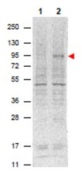
- Western blot using AM08449PU STAT5 pTyr694 antibody shows detection of phosphorylated Stat5 (indicated by arrowhead at ~91 kDa) in NK92 cells after 30 min treatment with 1Ku of IL-2 (Lane 2). No reactivity is seen for non-phosphorylated Stat5 in untreated cells (Lane 1). The membrane was probed with the primary antibody at a 1/1,000 dilution, overnight at 4°C. For detection DyLightâ¢800 conjugated Goat anti-Mouse IgG was used at a 1/20,000 dilution for 30 min at RT followed by visualization using a VersaDoc⢠MP 4000 imaging system (Bio-Rad).

Supportive validation
- Submitted by
- Acris Antibodies GmbH (provider)
- Main image
- Experimental details
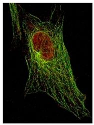
- AM08449PU-N STAT5 pTyr694 antibody staining of 3T3 cells by Immunofluorescent STED Microscopy. Red represents Stat5 pTyr694 protein. Green represents Tubulin.

Supportive validation
- Submitted by
- Acris Antibodies GmbH (provider)
- Main image
- Experimental details
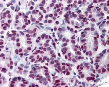
- Immunohistochemistry using AM08449PU STAT5 pTyr694 antibody shows detection of phosphorylated STAT5 pTyr694 in Human breast tissue (40X). The antibody was used a dilution to 20 μg/mL. The image shows breast epithelium with moderate nuclear staining. Tissue was Formalin Fixed, Paraffin Embedded. No pre-treatment of sample was required. The image shows the localization of antibody as the precipitated red signal, with a hematoxylin purple nuclear counterstain.Personal communication, Andrew Elston, Lifespan Biosciences, Seattle, WA.