Explore
 Validate
 Learn
Learn Western blot
Antibody data
- Antibody Data
- Antigen structure
- References [5]
- Comments [0]
- Validations
- Western blot [1]
- Immunohistochemistry [1]
Submit
Validation data
Reference
Comment
Report error
- Product number
- NB100-56558 - Provider product page

- Provider
- Novus Biologicals
- Proper citation
- Novus Cat#NB100-56558, RRID:AB_838374
- Product name
- Mouse Monoclonal HTRA2/Omi Antibody
- Antibody type
- Monoclonal
- Description
- Protein G purified. This antibody does not cross-react with mouse HtrA2.
- Reactivity
- Human
- Host
- Mouse
- Isotype
- IgG
- Vial size
- 0.1 mg
- Storage
- Store at 4C short term. Aliquot and store at -20C long term. Avoid freeze-thaw cycles.
Submitted references Apoptotic death concurrent with CD3 stimulation in primary human CD8+ T lymphocytes: a role for endogenous granzyme B.
Apoptotic death concurrent with CD3 stimulation in primary human CD8+ T lymphocytes: a role for endogenous granzyme B.
X-linked inhibitor of apoptosis (XIAP) protein protects against caspase activation and tissue loss after neonatal hypoxia-ischemia.
Immunohistochemical analysis of Omi/HtrA2 expression in stomach cancer.
Immunohistochemical analysis of Omi/HtrA2 expression in stomach cancer.
Laforge M, Bidère N, Carmona S, Devocelle A, Charpentier B, Senik A
Journal of immunology (Baltimore, Md. : 1950) 2006 Apr 1;176(7):3966-77
Journal of immunology (Baltimore, Md. : 1950) 2006 Apr 1;176(7):3966-77
Apoptotic death concurrent with CD3 stimulation in primary human CD8+ T lymphocytes: a role for endogenous granzyme B.
Laforge M, Bidère N, Carmona S, Devocelle A, Charpentier B, Senik A
Journal of immunology (Baltimore, Md. : 1950) 2006 Apr 1;176(7):3966-77
Journal of immunology (Baltimore, Md. : 1950) 2006 Apr 1;176(7):3966-77
X-linked inhibitor of apoptosis (XIAP) protein protects against caspase activation and tissue loss after neonatal hypoxia-ischemia.
Wang X, Zhu C, Wang X, Hagberg H, Korhonen L, Sandberg M, Lindholm D, Blomgren K
Neurobiology of disease 2004 Jun;16(1):179-89
Neurobiology of disease 2004 Jun;16(1):179-89
Immunohistochemical analysis of Omi/HtrA2 expression in stomach cancer.
Lee SH, Lee JW, Kim HS, Kim SY, Park WS, Kim SH, Lee JY, Yoo NJ
APMIS : acta pathologica, microbiologica, et immunologica Scandinavica 2003 May;111(5):586-90
APMIS : acta pathologica, microbiologica, et immunologica Scandinavica 2003 May;111(5):586-90
No comments: Submit comment
Supportive validation
- Submitted by
- Novus Biologicals (provider)
- Main image
- Experimental details
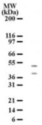
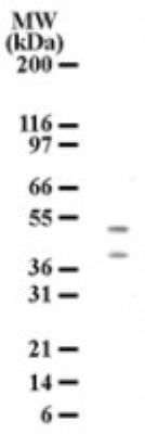
- Western Blot: HTRA2/Omi Antibody (196C429) [NB100-56558] - Detection of HtrA2 using an HtrA2 monoclonal antibody. HL-60 cell lysate was probed with HtrA2 antibody at 3 ug/ml and two protein bands of approximate molecular weight of 50 and 38 kDa were detected. The 50 kDa and 38 kDa bands may represent precursor and mature forms of HtrA2.
Supportive validation
- Submitted by
- Novus Biologicals (provider)
- Main image

- Experimental details
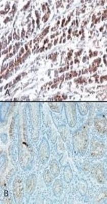
- Immunohistochemistry: HTRA2/Omi Antibody (196C429) [NB100-56558] - Analysis of HtrA2 using an HtrA2 monoclonal antibody. Probing with HtrA2 antibody shows staining in stomach tumor tissue (A) and very weak staining in normal stomach tissue (B).