Explore
 Validate
 Learn
Learn Western blot
 ELISA
ELISA Immunocytochemistry
Antibody data
- Antibody Data
- Antigen structure
- References [1]
- Comments [0]
- Validations
- Immunocytochemistry [5]
- Immunohistochemistry [1]
- Flow cytometry [3]
Submit
Validation data
Reference
Comment
Report error
- Product number
- 700238 - Provider product page

- Provider
- Invitrogen Antibodies
- Product name
- Phospho-4EBP1 (Thr37) Recombinant Rabbit Monoclonal Antibody (52H37L2)
- Antibody type
- Monoclonal
- Antigen
- Synthetic peptide
- Description
- This antibody is predicted to react with bovine, equine, mouse, opossum, rat and zebrafish based on sequence homology. Intact IgG appears on a non-reducing gel as ~150 kDa band and upon reduction generating a ~25 kDa light chain band and a ~50 kDa heavy chain. Recombinant rabbit monoclonal antibodies are produced using in vitro expression systems. The expression systems are developed by cloning in the specific antibody DNA sequences from immunoreactive rabbits. Then, individual clones are screened to select the best candidates for production. The advantages of using recombinant rabbit monoclonal antibodies include: better specificity and sensitivity, lot-to-lot consistency, animal origin-free formulations, and broader immunoreactivity to diverse targets due to larger rabbit immune repertoire.
- Reactivity
- Human
- Host
- Rabbit
- Isotype
- IgG
- Antibody clone number
- 52H37L2
- Vial size
- 100 μg
- Concentration
- 0.5 mg/mL
- Storage
- Store at 4°C short term. For long term storage, store at -20°C, avoiding freeze/thaw cycles.
Submitted references Sinoporphyrin sodium is a promising sensitizer for photodynamic and sonodynamic therapy in glioma.
An YW, Liu HQ, Zhou ZQ, Wang JC, Jiang GY, Li ZW, Wang F, Jin HT
Oncology reports 2020 Oct;44(4):1596-1604
Oncology reports 2020 Oct;44(4):1596-1604
No comments: Submit comment
Supportive validation
- Submitted by
- Invitrogen Antibodies (provider)
- Main image
- Experimental details
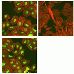
- Immunofluorescent analysis of Phospho-4EBP1 pThr37 in HeLa cells using a Phospho-4EBP1 pThr37 recombinant rabbit monoclonal antibody (Product # 700238) at a dilution of 2.5 µg/mL in the absence of peptide (top left) and presence of phosphopeptide used as immunogen (top right) or non-phosphopeptide (bottom left), followed by detection using an Alexa Fluor 488-conjugated goat anti-rabbit secondary antibody at a dilution of 1:1000. Actin was stained with Alexa Fluor 568 phalloidin (Product # A12380).

- Submitted by
- Invitrogen Antibodies (provider)
- Main image

- Experimental details
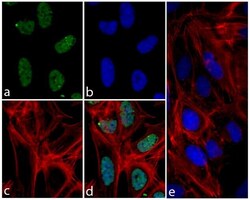
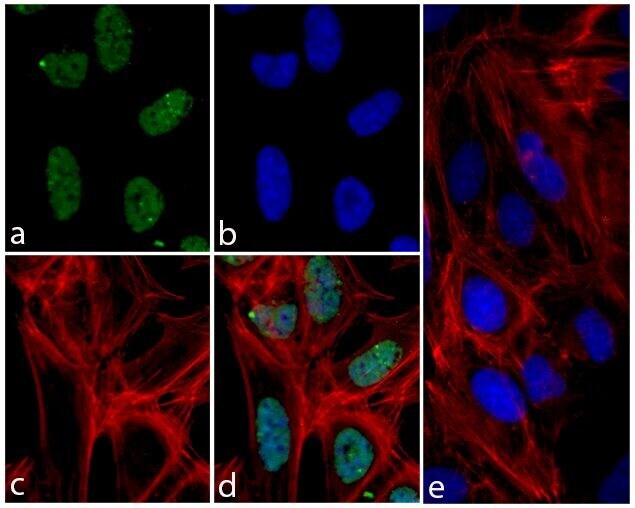
- Immunofluorescent analysis of 4E-BP1 (pT37) was done on 70% confluent log phase U2OS cells. The cells were fixed with 4% paraformaldehyde for 15 minutes; permeabilized with 0.25% Triton X-100 for 10 minutes followed by blocking with 5% BSA for 1 hour at room temperature. The cells were incubated with 4E-BP1 (pT37) Recombinant Rabbit Monoclonal Antibody (Product # 700238) at 2 µg-4 µg in 1% BSA and incubated for 3 hours at room temperature and then labeled with Alexa Fluor® 488 Goat anti-Rabbit IgG Secondary Antibody (Product # A-11008) at a dilution of 1:400 for 30 minutes at room temperature (Panel a: green). Nuclei (Panel b: blue) were stained with SlowFade® Gold Antifade Mountant with DAPI (Product # S36938). F-actin (Panel c: red) was stained with Alexa Fluor® 594 Phalloidin (Product # A12381). Panel d is a merged image showing nuclear localization of 4E-BP1 (pT37). Panel e shows competition with 4E-BP1 (pT37) peptide. The images were captured at 20X magnification.

- Submitted by
- Invitrogen Antibodies (provider)
- Main image

- Experimental details
- Immunofluorescent analysis of Phospho-4EBP1 pThr37 in HeLa cells using a Phospho-4EBP1 pThr37 recombinant rabbit monoclonal antibody (Product # 700238) at a dilution of 2.5 µg/mL in the absence of peptide (top left) and presence of phosphopeptide used as immunogen (top right) or non-phosphopeptide (bottom left), followed by detection using an Alexa Fluor 488-conjugated goat anti-rabbit secondary antibody at a dilution of 1:1000. Actin was stained with Alexa Fluor 568 phalloidin (Product # A12380).

- Submitted by
- Invitrogen Antibodies (provider)
- Main image
- Experimental details
- Immunofluorescent analysis of 4E-BP1 (pT37) was done on 70% confluent log phase U2OS cells. The cells were fixed with 4% paraformaldehyde for 15 minutes; permeabilized with 0.25% Triton X-100 for 10 minutes followed by blocking with 5% BSA for 1 hour at room temperature. The cells were incubated with 4E-BP1 (pT37) Recombinant Rabbit Monoclonal Antibody (Product # 700238) at 2 µg-4 µg in 1% BSA and incubated for 3 hours at room temperature and then labeled with Alexa Fluor® 488 Goat anti-Rabbit IgG Secondary Antibody (Product # A-11008) at a dilution of 1:400 for 30 minutes at room temperature (Panel a: green). Nuclei (Panel b: blue) were stained with SlowFade® Gold Antifade Mountant with DAPI (Product # S36938). F-actin (Panel c: red) was stained with Alexa Fluor® 594 Phalloidin (Product # A12381). Panel d is a merged image showing nuclear localization of 4E-BP1 (pT37). Panel e shows competition with 4E-BP1 (pT37) peptide. The images were captured at 20X magnification.
Supportive validation
- Submitted by
- Invitrogen Antibodies (provider)
- Main image
- Experimental details
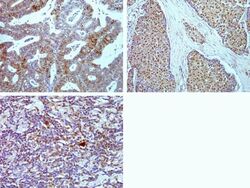
- Immunohistochemistry analysis of Phospho-4EBP1 pThr37 in formalin-fixed, paraffin-embedded human endometrial carcinoma (top left) breast carcinoma (top right) and Hodgkin lymphoma (bottom) using a Phospho-4EBP1 pThr37 monoclonal antibody (Product # 700238) at a dilution of 5 µg/mL. Images were taken at a magnification of 20x (top) or 40x (bottom). Results show strong nuclear staining in tumor cells.

Supportive validation
- Submitted by
- Invitrogen Antibodies (provider)
- Main image

- Experimental details
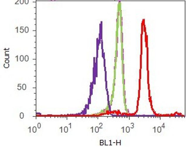
- Flow cytometry analysis of 4E-BP1 [pT37] was done on HeLa cells. Cells were fixed with 70% ethanol for 10 minutes, permeabilized with 0.25% Tritonª X-100 for 20 minutes, and blocked with 5% BSA for 1 hour at room temperature. Cells were labeled with ABfinityª 4E-BP1 [pT37] Recombinant Rabbit Monoclonal Antibody (700238, red histogram) or with rabbit isotype control (pink histogram) at 2 µg-4 µg/million cells in 2.5% BSA. After incubation at room temperature for 2-3 hours, the cells were labeled with Alexa Fluor¨ 488 Goat Anti-Rabbit Secondary Antibody (A11008) at a dilution of 1:400 for 30 minutes at room temperature. The representative 10,000 cells were acquired and analyzed for each sample using an Attune¨ Acoustic Focusing Cytometer. The purple histogram represents unstained control cells and the green histogram represents no-primary-antibody control.
- Submitted by
- Invitrogen Antibodies (provider)
- Main image
- Experimental details
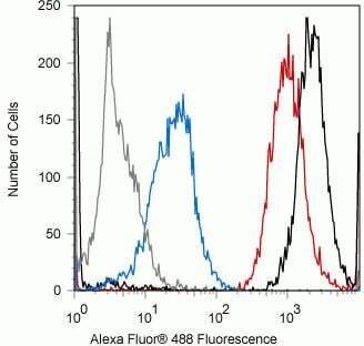
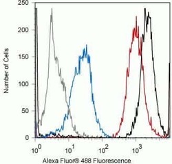
- Flow cytometry analysis of Phospho-4EBP1 pThr37 in Jurkat cells using a Phospho-4EBP1 pThr37 recombinant rabbit monoclonal antibody (Product # 700238) at a dilution of 0.1 µg. Cells were fixed and permeabilized using FIX & PERM (Product # GAS-004) reagent, and detection was performed using an Alexa Fluor 488 goat anti-rabbit IgG (black) compared to a control without primary antibody (gray). Pre-incubation with the phosphopeptide decreased the signal (blue) whereas incubation with the non-phosphopeptide did not (red).
- Submitted by
- Invitrogen Antibodies (provider)
- Main image

- Experimental details
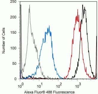
- Flow cytometry analysis of Phospho-4EBP1 pThr37 in Jurkat cells using a Phospho-4EBP1 pThr37 recombinant rabbit monoclonal antibody (Product # 700238) at a dilution of 0.1 µg. Cells were fixed and permeabilized using FIX & PERM (Product # GAS-004) reagent, and detection was performed using an Alexa Fluor 488 goat anti-rabbit IgG (black) compared to a control without primary antibody (gray). Pre-incubation with the phosphopeptide decreased the signal (blue) whereas incubation with the non-phosphopeptide did not (red).