Explore
 Validate
 Learn
Learn Western blot
 Immunocytochemistry
 Immunohistochemistry
Antibody data
- Antibody Data
- Antigen structure
- References [4]
- Comments [0]
- Validations
- Immunocytochemistry [6]
- Immunoprecipitation [1]
- Other assay [4]
Submit
Validation data
Reference
Comment
Report error
- Product number
- MA1-19331 - Provider product page

- Provider
- Invitrogen Antibodies
- Product name
- Fyn Monoclonal Antibody (FYN-01)
- Antibody type
- Monoclonal
- Antigen
- Recombinant protein fragment
- Description
- This antibody recognizes Fyn, a 59 kDa non-receptor Src-family protein tyrosine kinase (intracellular antigen). Immunoprecipitation: Preparation of cell lysate: 30 min on ice (orbital incubator) in lysing buffer, lysing buffer with N-dodecyl beta-D-maltoside (20 mM Tris/Cl, 100 mM NaCl pH 8,2, 1% laurylmaltosid (w/v), 50 mM NaF). The antibody FYN-01 apparently gives very specific signal (by 59 kDa), and nicely immunoprecipitants Fyn from cell lysates.
- Reactivity
- Human, Mouse
- Host
- Mouse
- Isotype
- IgG
- Antibody clone number
- FYN-01
- Vial size
- 100 μg
- Concentration
- 1 mg/mL
- Storage
- 4°C, do not freeze
Submitted references Human CEACAM1-LF regulates lipid storage in HepG2 cells via fatty acid transporter CD36.
A brain proteomic signature of incipient Alzheimer's disease in young APOE ε4 carriers identifies novel drug targets.
Fyn-tau Ablation Modifies PTZ-Induced Seizures and Post-seizure Hallmarks of Early Epileptogenesis.
Oxidation of KCNB1 potassium channels triggers apoptotic integrin signaling in the brain.
Chean J, Chen CJ, Gugiu G, Wong P, Cha S, Li H, Nguyen T, Bhatticharya S, Shively JE
The Journal of biological chemistry 2021 Nov;297(5):101311
The Journal of biological chemistry 2021 Nov;297(5):101311
A brain proteomic signature of incipient Alzheimer's disease in young APOE ε4 carriers identifies novel drug targets.
Roberts JA, Varma VR, An Y, Varma S, Candia J, Fantoni G, Tiwari V, Anerillas C, Williamson A, Saito A, Loeffler T, Schilcher I, Moaddel R, Khadeer M, Lovett J, Tanaka T, Pletnikova O, Troncoso JC, Bennett DA, Albert MS, Yu K, Niu M, Haroutunian V, Zhang B, Peng J, Croteau DL, Resnick SM, Gorospe M, Bohr VA, Ferrucci L, Thambisetty M
Science advances 2021 Nov 12;7(46):eabi8178
Science advances 2021 Nov 12;7(46):eabi8178
Fyn-tau Ablation Modifies PTZ-Induced Seizures and Post-seizure Hallmarks of Early Epileptogenesis.
Putra M, Puttachary S, Liu G, Lee G, Thippeswamy T
Frontiers in cellular neuroscience 2020;14:592374
Frontiers in cellular neuroscience 2020;14:592374
Oxidation of KCNB1 potassium channels triggers apoptotic integrin signaling in the brain.
Yu W, Gowda M, Sharad Y, Singh SA, Sesti F
Cell death & disease 2017 Apr 6;8(4):e2737
Cell death & disease 2017 Apr 6;8(4):e2737
No comments: Submit comment
Supportive validation
- Submitted by
- Invitrogen Antibodies (provider)
- Main image
- Experimental details
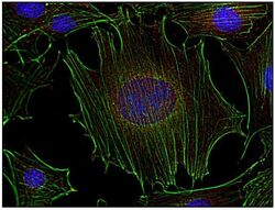
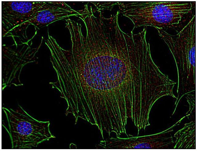
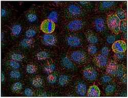
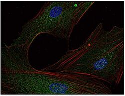
- Immunocytochemistry analysis of Fyn in murine transformed fibroblasts using anti-Fyn (FYN-01; red) Monoclonal antibody (Product # MA1-19331). Actin cytoskeleton was decorated by phalloidin (green) and cell nuclei stained with DAPI (blue).
- Submitted by
- Invitrogen Antibodies (provider)
- Main image
- Experimental details
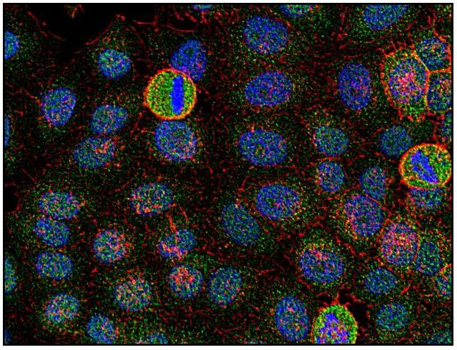
- Immunocytochemistry analysis of Fyn in human HeLa cell line using anti-Fyn (FYN-01; green) Monoclonal antibody (Product # MA1-19331). Actin cytoskeleton was decorated by phalloidin (red) and cell nuclei stained with DAPI (blue).

- Submitted by
- Invitrogen Antibodies (provider)
- Main image

- Experimental details
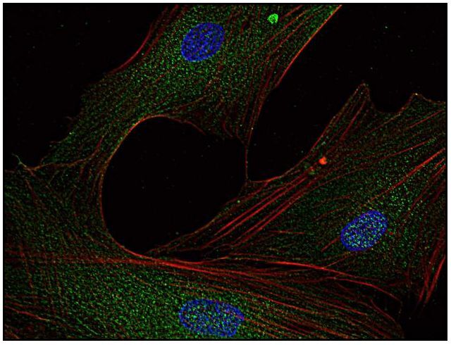
- Immunocytochemistry analysis of Fyn in human primary fibroblasts using anti-Fyn (FYN-01; green) Monoclonal antibody (Product # MA1-19331) using a dilution of 5 µg/mL. Actin cytoskeleton was decorated by phalloidin (red) and cell nuclei stained with DAPI (blue).

- Submitted by
- Invitrogen Antibodies (provider)
- Main image

- Experimental details
- Immunocytochemistry analysis of Fyn in murine transformed fibroblasts using anti-Fyn (FYN-01; red) Monoclonal antibody (Product # MA1-19331). Actin cytoskeleton was decorated by phalloidin (green) and cell nuclei stained with DAPI (blue).

- Submitted by
- Invitrogen Antibodies (provider)
- Main image

- Experimental details
- Immunocytochemistry analysis of Fyn in human HeLa cell line using anti-Fyn (FYN-01; green) Monoclonal antibody (Product # MA1-19331). Actin cytoskeleton was decorated by phalloidin (red) and cell nuclei stained with DAPI (blue).
- Submitted by
- Invitrogen Antibodies (provider)
- Main image
- Experimental details
- Immunocytochemistry analysis of Fyn in human primary fibroblasts using anti-Fyn (FYN-01; green) Monoclonal antibody (Product # MA1-19331) using a dilution of 5 µg/mL. Actin cytoskeleton was decorated by phalloidin (red) and cell nuclei stained with DAPI (blue).
Supportive validation
- Submitted by
- Invitrogen Antibodies (provider)
- Main image
- Experimental details
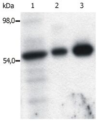
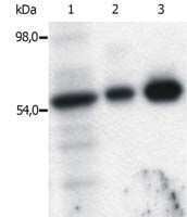
- Immunoprecipitation of Fyn from the lysate of T cells isolated from fresh buffy coats. Western blot was immunostained with anti-Fyn (FYN-01) Monoclonal antibody (Product # MA1-19331). Lane 1: original lysate of T cells; Lane 2-3: Immunoprecipitated material eluted from affinity sorbent (FYN-01 coupled to Sepharose beads). Lanes differ in amount of T cell lysate loaded on the immunosorbent.

Supportive validation
- Submitted by
- Invitrogen Antibodies (provider)
- Main image

- Experimental details
- Immunoprecipitation of Fyn using a monoclonal antibody (Product # MA1-19331).
- Submitted by
- Invitrogen Antibodies (provider)
- Main image

- Experimental details
- Immunoprecipitation of Fyn from the lysate of T cells isolated from fresh buffy coats. Western blot was immunostained with anti-Fyn (FYN-01) Monoclonal antibody (Product # MA1-19331). Lane 1: original lysate of T cells; Lane 2-3: Immunoprecipitated material eluted from affinity sorbent (FYN-01 coupled to Sepharose beads). Lanes differ in amount of T cell lysate loaded on the immunosorbent.

- Submitted by
- Invitrogen Antibodies (provider)
- Main image
- Experimental details
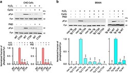
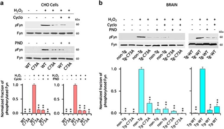
- Figure 6 Cyclo and PND-1186 inhibit Fyn activation induced by oxidation of KCNB1. ( a ) Representative western blots showing phosphorylated Fyn at tyr530 (pFyn) and total Fyn (Fyn) in CHO cells transfected with WT or C73A in the absence/presence of the indicated inhibitors. Fyn protein was detected into a single, ~60 kDa band. Cells were oxidized with 1.0 mM H 2 O 2 for 5 min and then incubated in control media or in media containing 200 nM Cyclo or 10 nM PND-1186 (PND) for 1 h before lysis. Quantifications of three experiments are shown in the lower panel and are normalized to WT+H 2 O 2 . P =1.3 x 10 -6 and 7.7 x 10 -7 for Cyclo and PND-1186, respectively (one-way analysis of variance (ANOVA)). ** P
- Submitted by
- Invitrogen Antibodies (provider)
- Main image

- Experimental details
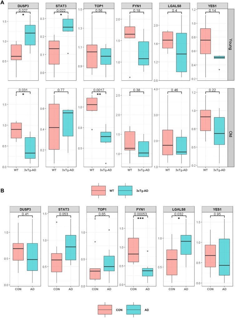
- Fig. 5. Primary validation of incipient AD proteomic signature. Results of primary validation analyses performed using Western blotting in 3xTg-AD transgenic mice (AD) compared to wild type (WT) ( A ) and human brain tissue samples comparing AD to CN ( B ). The 3xTg-AD ( n = 6) and WT ( n = 6) mice included young (38 to 40 weeks) and old (83 to 136 weeks) mice. (A) Box plots represent differences in protein levels in brain homogenates between 3xTg-AD (blue) and WT (red) mice. The top and bottom rows indicate protein levels in young and old mice, respectively. (B) Box plots represent differences in protein levels in brain homogenates between AD (blue; n = 11) and CN (red; n = 11). Protein levels that were significantly different between 3xTg-AD and WT or AD and CN are indicated with * ( P < 0.05) or ** ( P < 0.01). 3xTg-AD, 3xTg-AD transgenic mouse model of AD; AD, Alzheimer's disease; WT, wild type; DUSP3, dual-specificity phosphate 3; STAT3, signal transducer and activator of transcription 3; TOP1, DNA topoisomerase I; FYN1, FYN proto-oncogene tyrosine kinase; LGALS8, galectin-8; YES1, YES proto-oncogene tyrosine kinase.