Explore
 Validate
 Learn
Learn Western blot
Antibody data
- Antibody Data
- Antigen structure
- References [0]
- Comments [0]
- Validations
- Western blot [1]
- Immunocytochemistry [1]
- Immunohistochemistry [1]
Submit
Validation data
Reference
Comment
Report error
- Product number
- 11541-100UG - Provider product page

- Provider
- Invitrogen Antibodies
- Product name
- TrpC5 Ca+2 Channel Monoclonal Antibody (S67-15)
- Antibody type
- Monoclonal
- Antigen
- Synthetic peptide
- Reactivity
- Human, Mouse, Rat
- Host
- Mouse
- Isotype
- IgG
- Antibody clone number
- S67-15
- Vial size
- 100 µg
- Concentration
- 0.5-1.0 mg/mL
- Storage
- 2-8°C
No comments: Submit comment
Supportive validation
- Submitted by
- Invitrogen Antibodies (provider)
- Main image
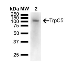
- Experimental details
- Western Blot analysis of Mouse brain showing detection of 110 kDa TrpC5 protein using Mouse Anti-TrpC5 Monoclonal Antibody, Clone N67/15 (11541). Lane 1: Molecular Weight Ladder (MW). Lane 2: Mouse Brain. Load: 15 ug. Block: 5% Skim Milk powder in TBST. Primary Antibody: Mouse Anti-TrpC5 Monoclonal Antibody (11541) at 1:1000 for Overnight at 4C. Secondary Antibody: Goat anti-mouse IgG:HRP at 1:7000 for 1 hour at RT with shaking. Color Development: Chemiluminescent for HRP (Moss) for 5 min in RT. Predicted/Observed Size: 110 kDa.

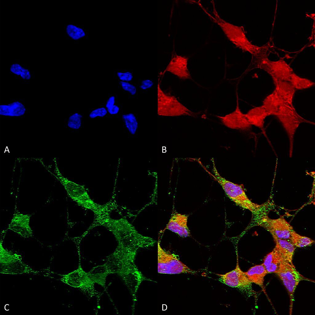
Supportive validation
- Submitted by
- Invitrogen Antibodies (provider)
- Main image
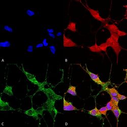
- Experimental details
- Immunocytochemistry/Immunofluorescence analysis using Mouse Anti-TrpC5 Monoclonal Antibody, Clone N67/15 (11541). Tissue: Neuroblastoma cells (SH-SY5Y). Species: Human. Fixation: 4% PFA for 15 min. Primary Antibody: Mouse Anti-TrpC5 Monoclonal Antibody (11541) at 1:50 for overnight at 4°C with slow rocking. Secondary Antibody: AlexaFluor 488 at 1:1000 for 1 hour at RT. Counterstain: Phalloidin-iFluor 647 (red) F-Actin stain; Hoechst (blue) nuclear stain at 1:800, 1.6mM for 20 min at RT. (A) Hoechst (blue) nuclear stain. (B) Phalloidin-iFluor 647 (red) F-Actin stain. (C) TrpC5 Antibody (D) Composite.
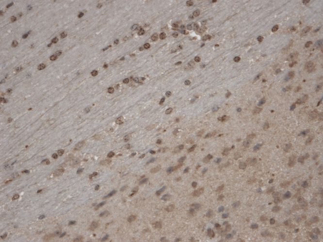
Supportive validation
- Submitted by
- Invitrogen Antibodies (provider)
- Main image
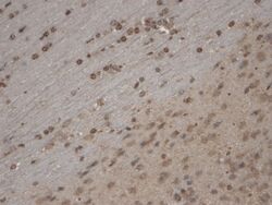
- Experimental details
- Immunohistochemistry analysis using Mouse Anti-TrpC5 Monoclonal Antibody, Clone N67/15 (11541). Tissue: Brain Slice. Species: Mouse. Fixation: 10% Formalin Solution for 12-24 hours at RT. Primary Antibody: Mouse Anti-TrpC5 Monoclonal Antibody (11541) at 1:1000 for 1 hour at RT. Secondary Antibody: HRP/DAB Detection System: Biotinylated Goat Anti-Mouse, Streptavidin Peroxidase, DAB Chromogen (brown) for 30 minutes at RT. Counterstain: Mayer Hematoxylin (purple/blue) nuclear stain at 250-500 µl for 5 minutes at RT. Localization: Nuclear staining. Magnification: 10X.