Explore
 Validate
 Learn
Learn Western blot
 ELISA
ELISA Immunocytochemistry
Antibody data
- Antibody Data
- Antigen structure
- References [2]
- Comments [0]
- Validations
- Immunocytochemistry [2]
- Immunohistochemistry [1]
- Flow cytometry [2]
- Other assay [1]
Submit
Validation data
Reference
Comment
Report error
- Product number
- MA5-17061 - Provider product page

- Provider
- Invitrogen Antibodies
- Product name
- CRP Monoclonal Antibody (1G1)
- Antibody type
- Monoclonal
- Antigen
- Purifed from natural sources
- Description
- MA5-17061 targets CRP in FACS, IHC, indirect ELISA, and WB applications and shows reactivity with Human samples. The MA5-17061 immunogen is purified recombinant fragment of human CRP expressed in E. Coli. MA5-17061 detects CRP which has a predicted molecular weight of approximately 25kDa.
- Reactivity
- Human
- Host
- Mouse
- Isotype
- IgG
- Antibody clone number
- 1G1
- Vial size
- 100 μL
- Concentration
- Conc. Not Determined
- Storage
- Store at 4°C short term. For long term storage, store at -20°C, avoiding freeze/thaw cycles.
Submitted references Bacillus anthracis Poly-γ-D-Glutamate Capsule Inhibits Opsonic Phagocytosis by Impeding Complement Activation.
Limitations of In Vivo Reprogramming to Dopaminergic Neurons via a Tricistronic Strategy.
Sharma S, Bhatnagar R, Gaur D
Frontiers in immunology 2020;11:462
Frontiers in immunology 2020;11:462
Limitations of In Vivo Reprogramming to Dopaminergic Neurons via a Tricistronic Strategy.
Theodorou M, Rauser B, Zhang J, Prakash N, Wurst W, Schick JA
Human gene therapy methods 2015 Aug;26(4):107-22
Human gene therapy methods 2015 Aug;26(4):107-22
No comments: Submit comment
Supportive validation
- Submitted by
- Invitrogen Antibodies (provider)
- Main image

- Experimental details
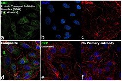
- Immunofluorescence analysis of C-reactive protein was performed using 70% confluent log phase Hep G2 cells. The cells were fixed with 4% paraformaldehyde for 10 minutes, permeabilized with 0.1% Triton™ X-100 for 15 minutes, and blocked with 2% BSA for 1 hour at room temperature. The cells were labeled with CRP Monoclonal Antibody (1G1) (Product # MA5-17061) at 1:200 dilution in 0.1% BSA, incubated at 4 degree celsius overnight and then labeled with Donkey anti-Mouse IgG (H+L) Highly Cross-Adsorbed Secondary Antibody, Alexa Fluor Plus 488 (Product # A32766, 1:2000 dilution), for 45 minutes at room temperature (Panel a: Green). Nuclei (Panel b:Blue) were stained with ProLong™ Diamond Antifade Mountant with DAPI (Product # P36962). F-actin (Panel c: Red) was stained with Rhodamine Phalloidin (Product # R415, 1:300 dilution). Panel d represents the merged image showing cytoplasmic localization. Panel e represents untreated HepG2 cells showing lower levels of CRP expression. Panel f represents control cells with no primary antibody to assess background. The images were captured at 60X magnification.

- Submitted by
- Invitrogen Antibodies (provider)
- Main image
- Experimental details
- Immunofluorescence analysis of C-reactive protein was performed using 70% confluent log phase Hep G2 cells. The cells were fixed with 4% paraformaldehyde for 10 minutes, permeabilized with 0.1% Triton™ X-100 for 15 minutes, and blocked with 2% BSA for 1 hour at room temperature. The cells were labeled with CRP Monoclonal Antibody (1G1) (Product # MA5-17061) at 1:200 dilution in 0.1% BSA, incubated at 4 degree celsius overnight and then labeled with Donkey anti-Mouse IgG (H+L) Highly Cross-Adsorbed Secondary Antibody, Alexa Fluor Plus 488 (Product # A32766, 1:2000 dilution), for 45 minutes at room temperature (Panel a: Green). Nuclei (Panel b:Blue) were stained with ProLong™ Diamond Antifade Mountant with DAPI (Product # P36962). F-actin (Panel c: Red) was stained with Rhodamine Phalloidin (Product # R415, 1:300 dilution). Panel d represents the merged image showing cytoplasmic localization. Panel e represents untreated HepG2 cells showing lower levels of CRP expression. Panel f represents control cells with no primary antibody to assess background. The images were captured at 60X magnification.

Supportive validation
- Submitted by
- Invitrogen Antibodies (provider)
- Main image

- Experimental details
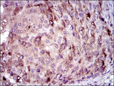
- Immunohistochemical analysis of paraffin-embedded liver cancer tissues using CRP monoclonal antibody (Product # MA5-17061) followed with DAB staining.
Supportive validation
- Submitted by
- Invitrogen Antibodies (provider)
- Main image

- Experimental details
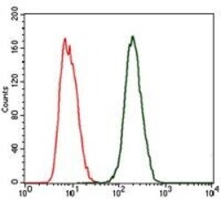
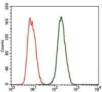
- Flow cytometric analysis of MCF-7 cells using CRP monoclonal antibody (Product # MA5-17061) (green) and negative control (red).

- Submitted by
- Invitrogen Antibodies (provider)
- Main image
- Experimental details
- Flow cytometric analysis of MCF-7 cells using CRP monoclonal antibody (Product # MA5-17061) (green) and negative control (red).
Supportive validation
- Submitted by
- Invitrogen Antibodies (provider)
- Main image
- Experimental details
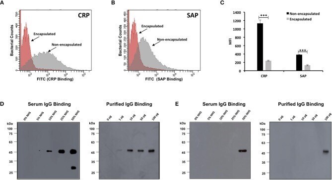
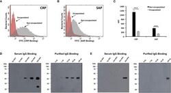
- Figure 5 Binding of complement mediators with encapsulated and non-encapsulated strains of Bacillus anthracis . Flow cytometry histogram for binding of pentraxins C-reactive protein (CRP) (A) and serum amyloid P component (SAP) (B) on B. anthracis strains incubated in 10% human serum. Gray and red shading indicates non-encapsulated and encapsulated bacteria, respectively. (C) CRP and SAP binding represented as mean fluorescence index (MFI) observed with non-encapsulated (black bars) and encapsulated bacteria (gray bars). Each bar represents the mean of three independent experiments. Error bars represent standard error of the mean. The scatter plots for the corresponding histograms are represented in Figures S4 , S5 . (D,E) Immunoblot assay for detection of human serum IgG and purified human IgG binding with B. anthracis non-encapsulated strains (D) and encapsulated strains (E) incubated with increasing concentration of normal human serum (5-50%) and purified human IgG (1-100 mug/ml); 1% normal human serum and 10 mug/ml of IgG was used as positive control. Statistical significance is highlighted by the following denotations: *** for P -value < 0.001.