 Explore
 Validate
 Learn
Learn Western blot
Antibody data
- Antibody Data
- Antigen structure
- References [0]
- Comments [0]
- Validations
- Western blot [2]
- Immunohistochemistry [1]
Submit
Validation data
Reference
Comment
Report error
- Product number
- 710269 - Provider product page

- Provider
- Invitrogen Antibodies
- Product name
- CRP Recombinant Polyclonal Antibody (5HCLC)
- Antibody type
- Polyclonal
- Antigen
- Recombinant full-length protein
- Description
- This antibody is predicted to react with non-human primate, rat and rabbit based on sequence homology.
- Antibody clone number
- 5HCLC
- Concentration
- 0.5 mg/mL
No comments: Submit comment
Supportive validation
- Submitted by
- Invitrogen Antibodies (provider)
- Main image

- Experimental details
- Western blot was performed using Anti-CRP Recombinant Polyclonal Antibody (5HCLC) (Product # 710269) and a 25 kDa band corresponding to C-reactive protein was observed in HepG2 upon IL-6 and PTI treatment. Whole cell extracts (60 µg lysate) of Hep G2 (Lane 1), Hep G2 (Protein Transport Inhibitor Cocktail (500X) (1X, 4 hours)) (Lane 2), Hep G2 (IL-6 and Protein Transport Inhibitor Cocktail (500X) (10 ng/mL, 1X; 24 hours,4 hours)) (Lane 3) were electrophoresed using NuPAGE™ 12% Bis-Tris Protein Gel (Product # NP0341BOX). Resolved proteins were then transferred onto a nitrocellulose membrane (Product # IB23001) by iBlot® 2 Dry Blotting System (Product # IB21001). The blot was probed with the primary antibody (1 µg/mL) and detected by chemiluminescence with Goat anti-Rabbit IgG (H+L) Superclonal™ Recombinant Secondary Antibody, HRP (Product # A27036,1:4000) using the iBright FL 1000 (Product # A32752). Chemiluminescent detection was performed using SuperSignal™ West Atto Ultimate Sensitivity Substrate (Product # A38556).

- Submitted by
- Invitrogen Antibodies (provider)
- Main image

- Experimental details
- Western blot analysis of CRP in whole cell extracts from NIH-3T3 cells using a CRP Recombinant Rabbit Polyclonal Antibody (Product # 710269) at a dilution of 1 µg/mL. Samples were detected using chemiluminescence (ECL). Results show a band at ~25kDa.

Supportive validation
- Submitted by
- Invitrogen Antibodies (provider)
- Main image
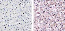
- Experimental details
- Immunohistochemistry analysis of C-Reactive Protein showing staining in the cytoplasm and weak staining in the nucleus of paraffin-embedded human liver tissue (right) compared to a negative control without primary antibody (left). To expose target proteins, antigen retrieval was performed using 10mM sodium citrate (pH 6.0), microwaved for 8-15 min. Following antigen retrieval, tissues were blocked in 3% H2O2-methanol for 15 min at room temperature, washed with ddH2O and PBS, and then probed with a C-Reactive Protein Recombinant Rabbit Polyclonal Antibody (Product # 710269) diluted in 3% BSA-PBS at a dilution of 1:20 for 1 hour at 37ºC in a humidified chamber. Tissues were washed extensively in PBST and detection was performed using an HRP-conjugated secondary antibody followed by colorimetric detection using a DAB kit. Tissues were counterstained with hematoxylin and dehydrated with ethanol and xylene to prep for mounting.
