 Explore
 Validate
 Learn
Learn ELISA
ELISA Immunocytochemistry
Antibody data
- Antibody Data
- Antigen structure
- References [0]
- Comments [0]
- Validations
- Immunocytochemistry [1]
Submit
Validation data
Reference
Comment
Report error
- Product number
- MA5-14744 - Provider product page

- Provider
- Invitrogen Antibodies
- Product name
- CRP Monoclonal Antibody (P4D7)
- Antibody type
- Monoclonal
- Antigen
- Purifed from natural sources
- Description
- MA5-14744 targets C Reactive Protein in ELISA, IP, and RIA applications and shows reactivity with Human samples. The MA5-14744 immunogen is purified human plasma CRP. MA5-14744 detects C Reactive Protein which has a predicted molecular weight of approximately 23 kDa. Product MA514744 is a smaller package size of MIC0501 (formerly sold as a Seradyn product).
- Reactivity
- Human
- Host
- Mouse
- Isotype
- IgG
- Antibody clone number
- P4D7
- Vial size
- 100 μg
- Concentration
- 1 mg/mL
- Storage
- Maintain refrigerated at 2-8°C for up to 6 months. For long term storage store at -20°C
No comments: Submit comment
Supportive validation
- Submitted by
- Invitrogen Antibodies (provider)
- Main image
- Experimental details
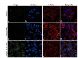
- Immunofluorescence analysis of CRP was performed using 70% confluent log phase HepG2 cells. The cells were fixed with 4% paraformaldehyde for 10 minutes, permeabilized with 0.1% Triton™ X-100 for 15 minutes, and blocked with 2% BSA for 45 minutes at room temperature. The cells were labeled with CRP Monoclonal Antibody (P4D7) (Product # MA5-14744, 5 µg/mL) in 0.1% BSA, incubated at 4 degree celsius overnight and then labeled with Donkey anti-Mouse IgG (H+L) Highly Cross-Adsorbed Secondary Antibody, Alexa Fluor Plus 488 (Product # A32766, 1:2000 dilution), for 45 minutes at room temperature (Panels a,e,i: Green). Nuclei (Panels b,f,j: Blue) were stained with ProLong™ Diamond Antifade Mountant with DAPI (Product # P36962). F-actin (Panels c,g,k: Red) was stained with Rhodamine Phalloidin (Product # R415, 1:300 dilution). Panels d,h,l represents the merged images showing cytoplasmic localization upon treatment with IL-6 and PTI. Panel (a-d) shows representative HepG2 control cells, whereas Panel (e-h) represent HepG2 with Protein Transport Inhibitor Cocktail (PTI)(500X) (1X, 4 hours) treatment. Similarly, panel (i-l) represent HepG2 cells treated with IL-6 and Protein Transport Inhibitor Cocktail (PTI)(500X) (10 ng/mL, 1X; 24 hours, 4 hours). The images were captured at 60X magnification.
