Explore
 Validate
 Learn
Learn Western blot
Antibody data
- Antibody Data
- Antigen structure
- References [0]
- Comments [0]
- Validations
- Western blot [1]
- Immunocytochemistry [1]
- Immunohistochemistry [1]
- Flow cytometry [1]
- Other assay [3]
Submit
Validation data
Reference
Comment
Report error
- Product number
- 44-1150G - Provider product page

- Provider
- Invitrogen Antibodies
- Product name
- Phospho-AMPK alpha-1,2 (Thr183, Thr172) Polyclonal Antibody
- Antibody type
- Polyclonal
- Antigen
- Synthetic peptide
- Reactivity
- Human, Mouse
- Host
- Rabbit
- Isotype
- IgG
- Vial size
- 100 µL
- Storage
- -20°C
No comments: Submit comment
Supportive validation
- Submitted by
- Invitrogen Antibodies (provider)
- Main image

- Experimental details
- Upregulation, Antibody-Peptide Competition and Phosphatase Stripping. Extracts of HepG2 cells untreated (1) or treated with 12 mM Metformin for 24 hours in serum free media (2-6) were resolved by SDS-PAGE on a 10% Tris-glycine gel and transferred to PVDF. The membrane was left untreated (1-5) or treated with lambda phosphatase (6), blocked with a 3% BSA-TBST buffer for one hour at room temperature, and then incubated with the Phospho-AMPK alpha-1,2 (Thr183, Thr172) antibody (Product # 44-1150G) for two hours at room temperature in 3% BSA-TBST buffer, following prior incubation with: no peptide (1,2), the non-phosphopeptide corresponding to the phosphopeptide immunogen (3), a generic phosphothreonine-containing peptide (4), or the phosphopeptide immunogen (5). After washing, the membrane was incubated with goat F(ab’)2 anti-rabbit IgG HRP conjugate (Product # ALI4404) and signals were detected using the Pierce SuperSignal™ method. The data show that only the phosphopeptide corresponding to Phospho-AMPK alpha-1,2 (Thr183, Thr172) completely blocks the signal and that phosphatase stripping eliminates the signal, verifying that the antibody is phosphorylation site-specific. The data also show upregulation of the phospho-signal upon Metformin treatment in this cell system.

Supportive validation
- Submitted by
- Invitrogen Antibodies (provider)
- Main image
- Experimental details
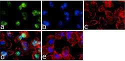
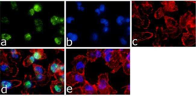
- Immunofluorescence analysis of Phospho-AMPK alpha 1,2 (Thr183, Thr172) was done on 70% confluent log phase MDA-MB-231 cells. The cells were fixed with 4% paraformaldehyde for 15 minutes, permeabilized with 0.25% Triton™ X-100 for 10 minutes, and blocked with 5% BSA for 1 hour at room temperature. The cells were labeled with Phospho-AMPK alpha-1,2 (Thr183, Thr172) Rabbit Polyclonal Antibody (Product # 44-1150G) at 1:250 dilution in 1% BSA and incubated for 3 hours at room temperature and then labeled with Goat anti-Rabbit IgG (H+L) Superclonal™ Secondary Antibody, Alexa Fluor® 488 conjugate (Product # A27034) at a dilution of 1:2000 for 45 minutes at room temperature (Panel a: green). Nuclei (Panel b: blue) were stained with SlowFade® Gold Antifade Mountant with DAPI (Product # S36938). F-actin (Panel c: red) was stained with Rhodamine Phalloidin (Product # R415, 1:300). Panel d is a merged image showing Nuclear localization. Panel e is a no primary antibody control. The images were captured at 60X magnification.
Supportive validation
- Submitted by
- Invitrogen Antibodies (provider)
- Main image

- Experimental details
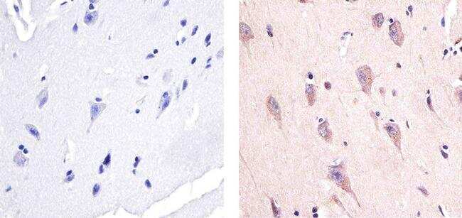
- Immunohistochemistry analysis of Phospho-AMPK alpha 1,2 (Thr183, Thr172) showing staining in the cytoplasm of paraffin-embedded human brain tissue (right) compared to a negative control without primary antibody (left). To expose target proteins, antigen retrieval was performed using 10mM sodium citrate (pH 6.0), microwaved for 8-15 min. Following antigen retrieval, tissues were blocked in 3% H2O2-methanol for 15 min at room temperature, washed with ddH2O and PBS, and then probed with a Phospho-AMPK alpha-1,2 (Thr183, Thr172) Rabbit Polyclonal Antibody (Product # 44-1150G) diluted in 3% BSA-PBS at a dilution of 1:20 overnight at 4°C in a humidified chamber. Tissues were washed extensively in PBST and detection was performed using an HRP-conjugated secondary antibody followed by colorimetric detection using a DAB kit. Tissues were counterstained with hematoxylin and dehydrated with ethanol and xylene to prep for mounting.
Supportive validation
- Submitted by
- Invitrogen Antibodies (provider)
- Main image
- Experimental details
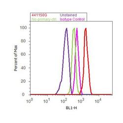
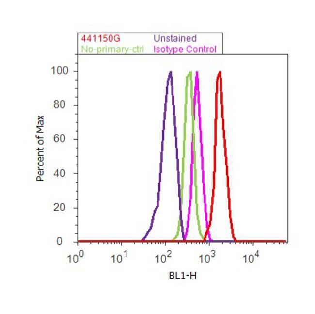
- Flow cytometry analysis of AMPK alpha 1 + 2 [pThr172] was done on MDA-MB-231 cells. Cells were fixed with 70% ethanol for 10 minutes, permeabilized with 0.25% Triton™ X-100 for 20 minutes, and blocked with 5% BSA for 30 minutes at room temperature. Cells were labeled with AMPK alpha 1 + 2 [pThr172] Rabbit Polyclonal Antibody (441150G, red histogram) or with rabbit isotype control (pink histogram) at 3-5 ug/million cells in 2.5% BSA. After incubation at room temperature for 2 hours, the cells were labeled with Alexa Fluor® 488 Goat Anti-Rabbit Secondary Antibody (A11008) at a dilution of 1:400 for 30 minutes at room temperature. The representative 10,000 cells were acquired and analyzed for each sample using an Attune® Acoustic Focusing Cytometer. The purple histogram represents unstained control cells and the green histogram represents no-primary-antibody control.
Supportive validation
- Submitted by
- Invitrogen Antibodies (provider)
- Main image

- Experimental details
- NULL
- Submitted by
- Invitrogen Antibodies (provider)
- Main image
- Experimental details
- NULL
- Submitted by
- Invitrogen Antibodies (provider)
- Main image

- Experimental details
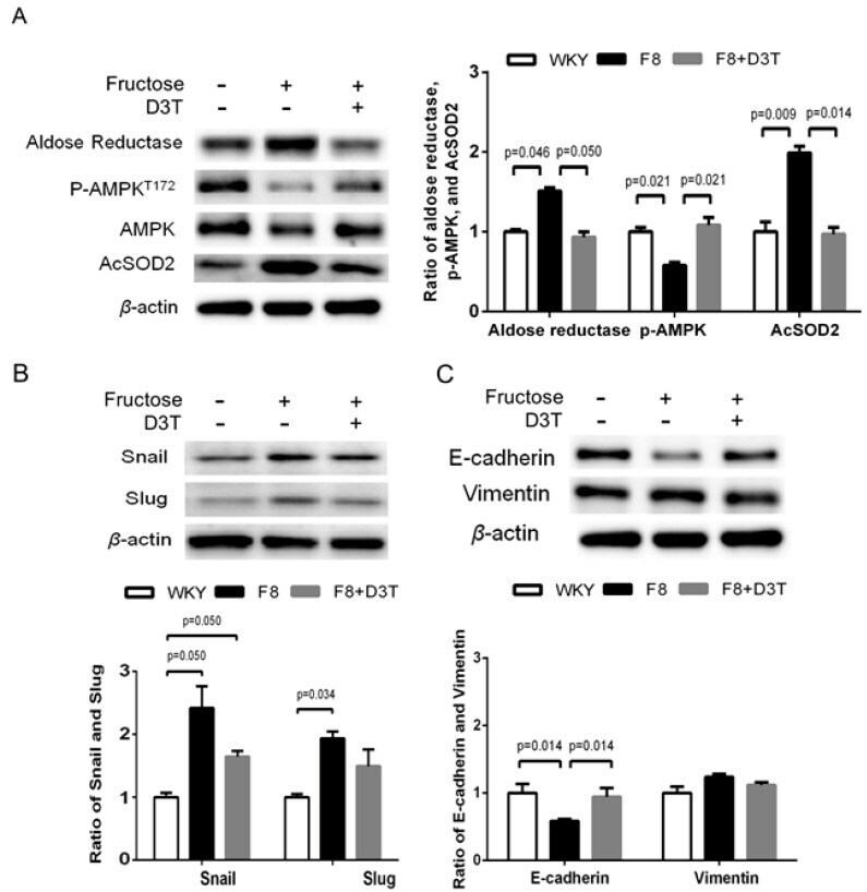
- Figure 4 D3T reduces fructose-induced EMT progression in the lens of rats with fructose-induced type 2 DM. ( A ) Immunoblotting analysis depicting Aldose reductase, p-AMPK T172 and Ac-SOD2 expression in the fructose-induced type 2 DM lens with or without D3T administration. ( B ) Immunoblotting analysis showing Snail and Slug protein expressions in the fruc Table 2 . DM lens with or without D3T administration. ( C ) Protein expressions of E-cadherin and vimentin were also analyzed and quantified. All data represented as mean +- SEM ( n = 6 per group, independent experiments in each figure).