 Explore
 Validate
 Learn
Learn Western blot
Antibody data
- Antibody Data
- Antigen structure
- References [0]
- Comments [0]
- Validations
- Western blot [3]
- Immunocytochemistry [1]
Submit
Validation data
Reference
Comment
Report error
- Product number
- 710099 - Provider product page

- Provider
- Invitrogen Antibodies
- Product name
- Phospho-AMPK alpha-1,2 (Thr183, Thr172) Recombinant Polyclonal Antibody (10HCLC)
- Antibody type
- Polyclonal
- Antigen
- Synthetic peptide
- Reactivity
- Human
- Host
- Rabbit
- Isotype
- IgG
- Antibody clone number
- 10HCLC
- Vial size
- 100 µg
- Concentration
- 0.5 mg/mL
- Storage
- Store at 4°C short term. For long term storage, store at -20°C, avoiding freeze/thaw cycles.
No comments: Submit comment
Supportive validation
- Submitted by
- Invitrogen Antibodies (provider)
- Main image

- Experimental details
- Western blot analysis was performed on whole cell extracts (30 µg lysate) of A-431 (Lane 1), A-431 treated with Metformin (10mM for 24 h) (Lane 2), Hep G2 (Lane 3) and Hep G2 treated with Metformin (10mM for 24 h) (Lane 4), U-87 MG (Lane 5), and U-87 MG treated with AICAR (1mM for 48 h) (Lane 6). The blot was probed with Phospho-AMPK alpha-1,2 (Thr183, Thr172) Monoclonal Antibody (Product # 710099, 2 µg/mL) and detected by chemiluminescence using Goat anti-Rabbit IgG (H+L) Superclonal™ Secondary Antibody, HRP conjugate (Product # A27036, 0.25 µg/mL, 1:4000 dilution). A 63 kDa band corresponding to Phospho-AMPK alpha-1 (Thr172) was observed across the cell lines tested and enhanced upon treatment.

- Submitted by
- Invitrogen Antibodies (provider)
- Main image
- Experimental details
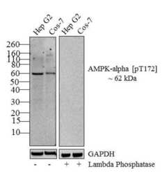
- Western blot analysis of Phospho-AMPK alpha (Thr183, Thr172) was performed by loading 20 µg of Hep G2 (lane1) and Cos-7 (lane2) lysate using Novex®NuPAGE® 4-12% Bis-Tris gel (Product # NP0321BOX), XCell SureLock Electrophoresis System (Product # EI0002), Novex® Sharp Pre-Stained Protein Standard (Product # LC5800), and iBlot® Dry Blotting System (Product # IB21001). Proteins were transferred to a nitrocellulose membrane and blocked with 5% skim milk for 1 hour at room temperature. Phospho-AMPK alpha (Thr183, ThrT172) was detected at ~ 62 kDa using Phospho-AMPK alpha-1,2 (Thr183, Thr172) Recombinant Rabbit Polyclonal Antibody (Product # 710099) at 1-3 µg/mL in 2.5% skim milk at 4°C overnight on a rocking platform. To confirm specificity, the corresponding blot on right was incubated with lambda phosphatase and its reactivity with antibody was tested. Goat anti-Rabbit IgG - HRP Secondary Antibody (Product # G-21234) at 1:5000 dilution was used and chemiluminescent detection was performed using Pierce™ ECL Western blotting Substrate (Product # 32106).

- Submitted by
- Invitrogen Antibodies (provider)
- Main image
- Experimental details
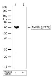
- Western blot analysis of Phospho-AMPK alpha (Thr183, Thr172) in whole cell extracts of HepG2 cells using a Phospho-AMPK alpha-1,2 (Thr183, Thr172) Recombinant Rabbit Polyclonal Antibody (Product # 710099) at a dilution of 2.5 µg/mL. To confirm specificity, competition was performed by preincubation with the phosphopeptide to inhibit antibody binding (lane 2). Results show a band at ~62kDa.

Supportive validation
- Submitted by
- Invitrogen Antibodies (provider)
- Main image
- Experimental details
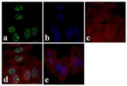
- Immunofluorescence analysis of Phospho-AMPK alpha (Thr183, Thr172) was done on 70% confluent log phase HeLa cells. The cells were fixed with 4% paraformaldehyde for 15 minutes, permeabilized with 0.25% Triton X-100 for 10 minutes, and blocked with 5% BSA for 1 hour at room temperature. The cells were labeled with Phospho-AMPK alpha-1,2 (Thr183, Thr172) Recombinant Rabbit Polyclonal Antibody (Product # 710099) at 3 in 1% BSA and incubated for 3 hours at room temperature and then labeled with Alexa Fluor 488 Goat anti-Rabbit IgG Secondary Antibody (Product # A-11008) at a dilution of 1:400 for 30 minutes at room temperature (Panel a: green). Nuclei (Panel b: blue) were stained with SlowFade® Gold Antifade Mountant DAPI (Product # S36938). F-actin (Panel c: red) was stained with Alexa Fluor 594 Phalloidin (Product # A12381). Panel d is a merged image showing nuclear localization. Panel e shows no primary antibody control. The images were captured at 20X magnification.
