Explore
 Validate
 Learn
Learn Western blot
 ELISA
ELISA Immunocytochemistry
Antibody data
- Antibody Data
- Antigen structure
- References [5]
- Comments [0]
- Validations
- Immunocytochemistry [6]
- Immunohistochemistry [1]
- Flow cytometry [2]
- Other assay [3]
Submit
Validation data
Reference
Comment
Report error
- Product number
- MA5-15815 - Provider product page

- Provider
- Invitrogen Antibodies
- Product name
- AMPK alpha-1 Monoclonal Antibody (2B7)
- Antibody type
- Monoclonal
- Antigen
- Purifed from natural sources
- Description
- MA5-15815 targets PRKAA1 in indirect ELISA, FACS, IF, IHC, and WB applications and shows reactivity with Human, mouse, Non-human primate, and Rat samples. The MA5-15815 immunogen is purified recombinant fragment of human PRKAA1 expressed in E. Coli. MA5-15815 detects PRKAA1 which has a predicted molecular weight of approximately 64kDa.
- Reactivity
- Human, Mouse, Rat
- Host
- Mouse
- Isotype
- IgG
- Antibody clone number
- 2B7
- Vial size
- 100 μL
- Concentration
- Conc. Not Determined
- Storage
- Store at 4°C short term. For long term storage, store at -20°C, avoiding freeze/thaw cycles.
Submitted references ACSL4 contributes to sevoflurane-induced ferroptotic neuronal death in SH-SY5Y cells via the 5' AMP-activated protein kinase/mammalian target of rapamycin pathway.
Antifungal itraconazole ameliorates experimental autoimmune encephalomyelitis through a novel mechanism of action.
α1AMP-Activated Protein Kinase Protects against Lipopolysaccharide-Induced Endothelial Barrier Disruption via Junctional Reinforcement and Activation of the p38 MAPK/HSP27 Pathway.
Rotating magnetic field delays human umbilical vein endothelial cell aging and prolongs the lifespan of Caenorhabditis elegans.
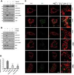
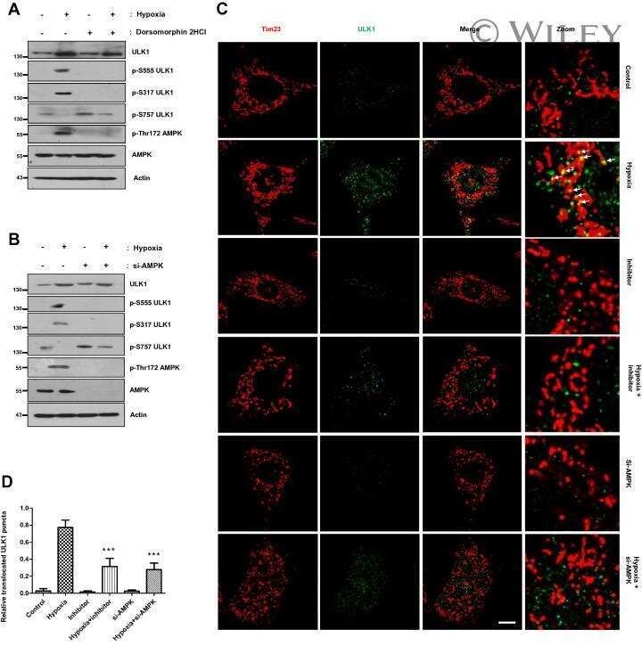
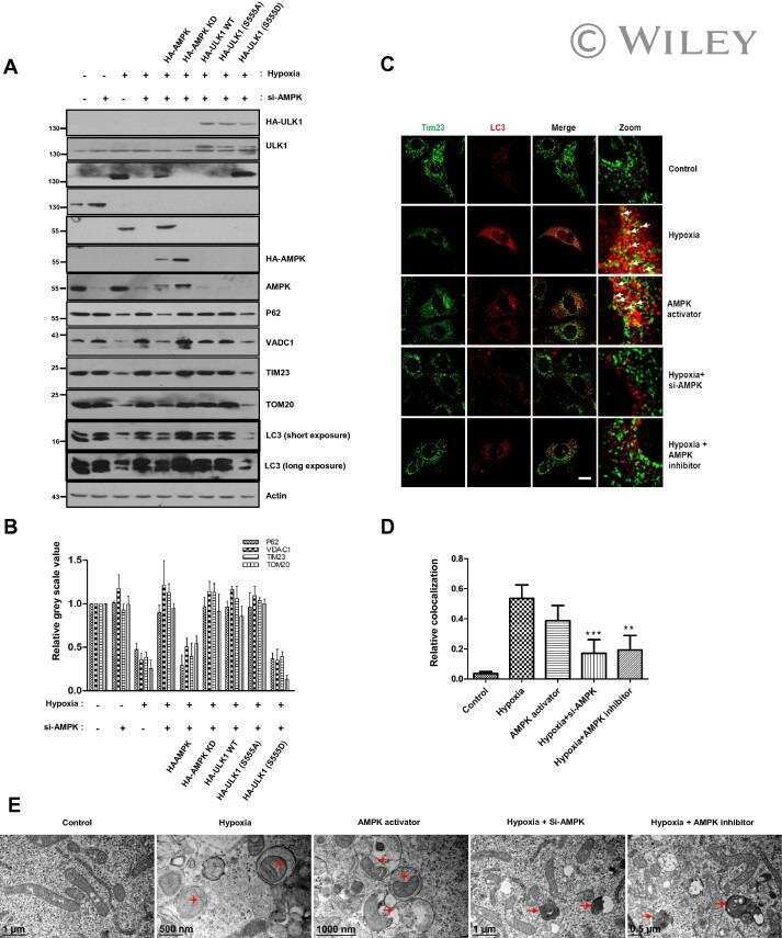
Phosphorylation of ULK1 by AMPK regulates translocation of ULK1 to mitochondria and mitophagy.
Cheng L, Zhu X, Liu Y, Zhu K, Lin K, Li F
Annals of translational medicine 2021 Sep;9(18):1454
Annals of translational medicine 2021 Sep;9(18):1454
Antifungal itraconazole ameliorates experimental autoimmune encephalomyelitis through a novel mechanism of action.
Huang H, Tian X, Peng X, Huang L, Mei L, Zhan Y, Chen S, Wu H, Wei G, Cai X
Advances in clinical and experimental medicine : official organ Wroclaw Medical University 2020 May;29(5):535-545
Advances in clinical and experimental medicine : official organ Wroclaw Medical University 2020 May;29(5):535-545
α1AMP-Activated Protein Kinase Protects against Lipopolysaccharide-Induced Endothelial Barrier Disruption via Junctional Reinforcement and Activation of the p38 MAPK/HSP27 Pathway.
Angé M, Castanares-Zapatero D, De Poortere J, Dufeys C, Courtoy GE, Bouzin C, Quarck R, Bertrand L, Beauloye C, Horman S
International journal of molecular sciences 2020 Aug 4;21(15)
International journal of molecular sciences 2020 Aug 4;21(15)
Rotating magnetic field delays human umbilical vein endothelial cell aging and prolongs the lifespan of Caenorhabditis elegans.
Xu J, Liu K, Chen T, Zhan T, Ouyang Z, Wang Y, Liu W, Zhang X, Sun Y, Xu G, Wang X
Aging 2019 Nov 22;11(22):10385-10408
Aging 2019 Nov 22;11(22):10385-10408
Phosphorylation of ULK1 by AMPK regulates translocation of ULK1 to mitochondria and mitophagy.
Tian W, Li W, Chen Y, Yan Z, Huang X, Zhuang H, Zhong W, Chen Y, Wu W, Lin C, Chen H, Hou X, Zhang L, Sui S, Zhao B, Hu Z, Li L, Feng D
FEBS letters 2015 Jul 8;589(15):1847-54
FEBS letters 2015 Jul 8;589(15):1847-54
No comments: Submit comment
Supportive validation
- Submitted by
- Invitrogen Antibodies (provider)
- Main image
- Experimental details
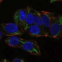
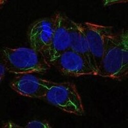
- Immunofluorescence analysis of NTERA-2 cells using PRKAA1 monoclonal antibody (Product # MA5-15815) (Green). Blue: DRAQ5 fluorescent DNA dye. Red: actin filaments have been labeled with phalloidin.

- Submitted by
- Invitrogen Antibodies (provider)
- Main image
- Experimental details
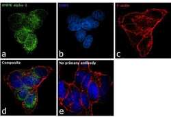
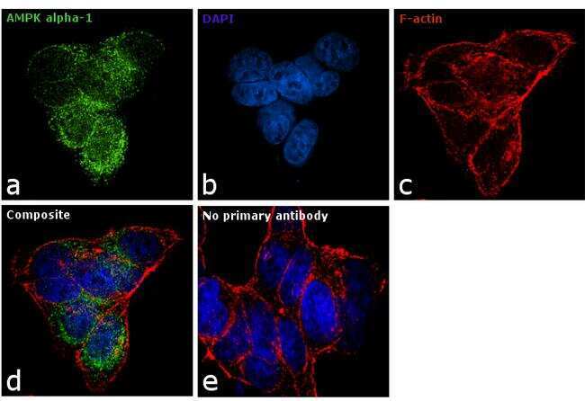
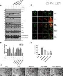
- Immunofluorescence analysis of AMPK alpha-1 was performed using 70% confluent log phase LNCap cells. The cells were fixed with 4% paraformaldehyde for 10 minutes, permeabilized with 0.1% Triton™ X-100 for 10 minutes, and blocked with 1% BSA for 1 hour at room temperature. The cells were labeled with AMPK alpha-1 Monoclonal Antibody (2B7)(Product # MA5-15815) at 1:100 dilution in 0.1% BSA and incubated overnight at 4 degree and then labeled with Goat anti-Mouse IgG (H+L) Superclonal™ Secondary Antibody, Alexa Fluor® 488 conjugate (A28175) at a dilution of 1:2000 for 45 minutes at room temperature (Panel a: green). Nuclei (Panel b: blue) were stained with SlowFade® Gold Antifade Mountant with DAPI (Product # S36938). F-actin (Panel c: red) was stained with Rhodamine Phalloidin (Product # R415, 1:300). Panel d represents the merged image showing mitochondrial localization. Panel e represents control cells with no primary antibody to assess background. The images were captured at 60X magnification.
- Submitted by
- Invitrogen Antibodies (provider)
- Main image

- Experimental details
- Immunofluorescence analysis of NTERA-2 cells using PRKAA1 monoclonal antibody (Product # MA5-15815) (Green). Blue: DRAQ5 fluorescent DNA dye. Red: actin filaments have been labeled with phalloidin.
- Submitted by
- Invitrogen Antibodies (provider)
- Main image
- Experimental details
- Immunofluorescence analysis of NTERA-2 cells using PRKAA1 monoclonal antibody (Product # MA5-15815) (Green). Blue: DRAQ5 fluorescent DNA dye. Red: actin filaments have been labeled with phalloidin.

- Submitted by
- Invitrogen Antibodies (provider)
- Main image

- Experimental details
- Immunofluorescence analysis of NTERA-2 cells using PRKAA1 monoclonal antibody (Product # MA5-15815) (Green). Blue: DRAQ5 fluorescent DNA dye. Red: actin filaments have been labeled with phalloidin.

- Submitted by
- Invitrogen Antibodies (provider)
- Main image

- Experimental details
- Immunofluorescence analysis of AMPK alpha-1 was performed using 70% confluent log phase LNCap cells. The cells were fixed with 4% paraformaldehyde for 10 minutes, permeabilized with 0.1% Triton™ X-100 for 10 minutes, and blocked with 1% BSA for 1 hour at room temperature. The cells were labeled with AMPK alpha-1 Monoclonal Antibody (2B7)(Product # MA5-15815) at 1:100 dilution in 0.1% BSA and incubated overnight at 4 degree and then labeled with Goat anti-Mouse IgG (H+L) Superclonal™ Secondary Antibody, Alexa Fluor® 488 conjugate (A28175) at a dilution of 1:2000 for 45 minutes at room temperature (Panel a: green). Nuclei (Panel b: blue) were stained with SlowFade® Gold Antifade Mountant with DAPI (Product # S36938). F-actin (Panel c: red) was stained with Rhodamine Phalloidin (Product # R415, 1:300). Panel d represents the merged image showing mitochondrial localization. Panel e represents control cells with no primary antibody to assess background. The images were captured at 60X magnification.

Supportive validation
- Submitted by
- Invitrogen Antibodies (provider)
- Main image
- Experimental details
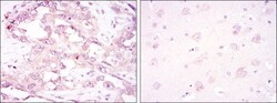
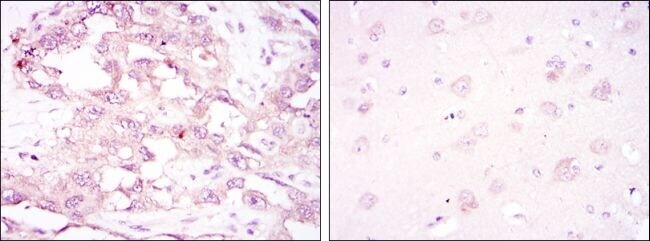
- Immunohistochemical analysis of paraffin-embedded ovarian cancer (left) and brain tissues (right) using PRKAA1 monoclonal antibody (Product # MA5-15815) followed with DAB staining.
Supportive validation
- Submitted by
- Invitrogen Antibodies (provider)
- Main image

- Experimental details
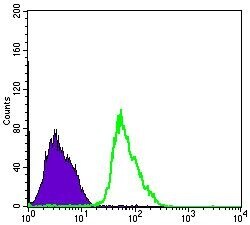
- Flow cytometric analysis of PC-2 cells using PRKAA1 monoclonal antibody (Product # MA5-15815) (green) and negative control (purple).
- Submitted by
- Invitrogen Antibodies (provider)
- Main image
- Experimental details
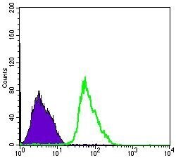
- Flow cytometric analysis of PC-2 cells using PRKAA1 monoclonal antibody (Product # MA5-15815) (green) and negative control (purple).

Supportive validation
- Submitted by
- Invitrogen Antibodies (provider)
- Main image
- Experimental details
- NULL
- Submitted by
- Invitrogen Antibodies (provider)
- Main image
- Experimental details
- NULL
- Submitted by
- Invitrogen Antibodies (provider)
- Main image
- Experimental details
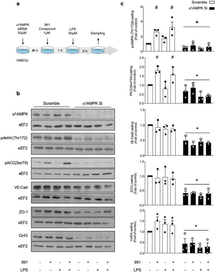
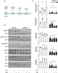
- Figure 1 Basal alpha1AMPK regulates VE-Cad, ZO-1, and Cx43 expression. ( a ) Schematic representation of the experimental design, in which HMECs were transfected with control non-targeting siRNA or alpha1AMPK siRNA (50 nM) for 48 h. Then, they were treated with 991 (1 muM) or DMSO for one hour and subsequently exposed or not exposed to lipopolysaccharide (LPS, 50 muM) for six hours; ( b , c ) Human dermal microvascular endothelial cells (HMECs) were treated according to the protocol detailed in ( a ). Cell lysates were submitted to Western blot analysis and probed with alpha1AMPK, phospho-AMPK (Thr172), phospho-ACC (Ser79), VE-Cad, ZO-1, and Cx43 antibodies (Abs). Anti-eukaryotic elongation factor 2 (eEF2) was used as a loading control. Representative western blots; ( b ) and quantifications ( c ) are shown. Data are expressed as mean +- standard deviation (SD) (three biological replicates for each condition). # p < 0.05 is relative to corresponding untreated HMECs and * p < 0.05 is relative to cells transfected with the scrambled siRNA. The data underwent two-way analysis of variance (ANOVA).