Antibody data
- Antibody Data
- Antigen structure
- References [1]
- Comments [0]
- Validations
- Immunocytochemistry [1]
- Immunohistochemistry [1]
- Other assay [1]
Submit
Validation data
Reference
Comment
Report error
- Product number
- PA5-29232 - Provider product page

- Provider
- Invitrogen Antibodies
- Product name
- NCK1 Polyclonal Antibody
- Antibody type
- Polyclonal
- Antigen
- Recombinant full-length protein
- Description
- Recommended positive controls: K562. Predicted reactivity: Mouse (98%), Rat (99%), Xenopus laevis (84%), Pig (97%), Sheep (98%), Rhesus Monkey (100%), Chimpanzee (100%), Bovine (99%). Store product as a concentrated solution. Centrifuge briefly prior to opening the vial.
- Reactivity
- Human
- Host
- Rabbit
- Isotype
- IgG
- Vial size
- 100 μL
- Concentration
- 0.78 mg/mL
- Storage
- Store at 4°C short term. For long term storage, store at -20°C, avoiding freeze/thaw cycles.
Submitted references Expression and clinicopathological significance of Nck1 in human astrocytoma progression.
Deshpande RP, Panigrahi M, Y B V K CS, Babu PP
The International journal of neuroscience 2019 Feb;129(2):171-178
The International journal of neuroscience 2019 Feb;129(2):171-178
No comments: Submit comment
Supportive validation
- Submitted by
- Invitrogen Antibodies (provider)
- Main image

- Experimental details
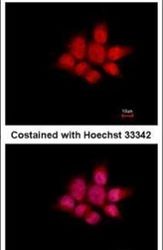
- Immunofluorescent analysis of NCK1 in paraformaldehyde-fixed A431 cells using a NCK1 polyclonal antibody (Product # PA5-29232) at a 1:500 dilution.
Supportive validation
- Submitted by
- Invitrogen Antibodies (provider)
- Main image

- Experimental details
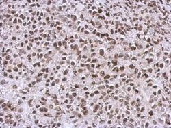
- Immunohistochemical analysis of paraffin-embedded Hela xenograft, using NCK1 (Product # PA5-29232) antibody at 1:500 dilution. Antigen Retrieval: EDTA based buffer, pH 8.0, 15 min.
Supportive validation
- Submitted by
- Invitrogen Antibodies (provider)
- Main image

- Experimental details
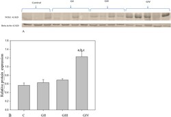
- Figure 2. Western blotting (A) Nck1 (1:1500 dilution) and densitometric analysis (B) of Nck1 in control, low-grade (GII) and high-grade (GIII, GIV) tumor tissue samples. Protein expression in GIV tissues was significantly ( p < 0.05) increased compared with control (a), GII (b), and GIII (c). Internal Nck1 expression were normalized with Beta actin (internal control) C = control brain tissue, GII = Grade-II astrocytoma tissue, GIII = Grade-III astrocytoma tissue, and GIV = Grade-IV astrocytoma tissue.
 Explore
 Validate
 Learn
Learn Western blot
 Immunocytochemistry