Explore
 Validate
 Learn
Learn Western blot
 ELISA
ELISAAntibody data
- Antibody Data
- Antigen structure
- References [0]
- Comments [0]
- Validations
- Western blot [1]
- Immunocytochemistry [1]
- Immunohistochemistry [1]
- Flow cytometry [1]
Submit
Validation data
Reference
Comment
Report error
- Product number
- GTX22806 - Provider product page

- Provider
- GeneTex
- Proper citation
- GeneTex Cat#GTX22806, RRID:AB_384862
- Product name
- ARF1/ARF3/ARF5/ARF6 antibody [1D9]
- Antibody type
- Monoclonal
- Reactivity
- Human, Mouse, Rat, Bovine, Canine, Hamster, Rabbit, Simian
- Host
- Mouse
- Storage
- -20¢X C, Avoid Freeze/Thaw Cycles
No comments: Submit comment
Supportive validation
- Submitted by
- GeneTex (provider)
- Main image

- Experimental details
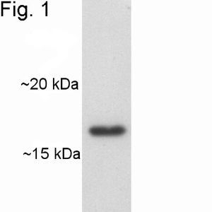
- Western blot of ADP Ribosylation Factor in canine heart extract.
Supportive validation
- Submitted by
- GeneTex (provider)
- Main image
- Experimental details
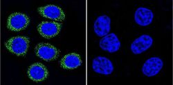
- Immunofluorescent analysis of ADP Ribosylation Factor in HeLa Cells. ADP Ribosylation Factor staining (green) and nuclei with DAPI (blue) is shown. Cells were grown on slides and fixed with formaldehyde prior to staining. Cells were probed without (control) or with ADP Ribosylation Factor antibody [1D9] at a dilution of 1:100 over night at 4 °C, washed with PBS and incubated with a proper secondary antibody. Images were taken at 60X magnification.

Supportive validation
- Submitted by
- GeneTex (provider)
- Main image
- Experimental details
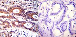
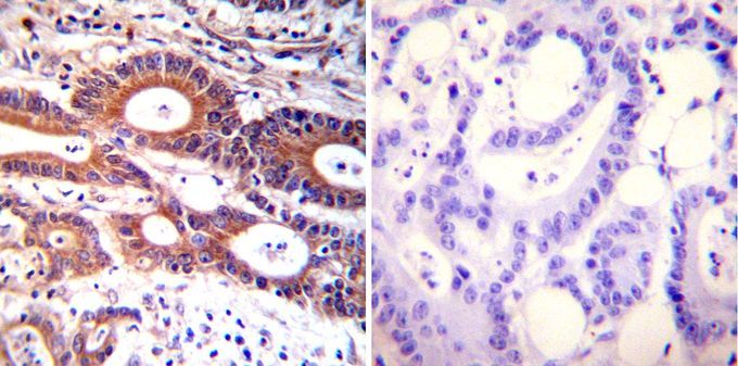
- Immunohistochemistry was performed on cancer biopsies of deparaffinized human colon carcinoma tissues. To expose target proteins, heat induced antigen retrieval was performed using 10mM sodium citrate (pH6.0) buffer, microwaved for 8-15 minutes. Following antigen retrieval tissues were blocked in 3% BSA-PBS for 30 minutes at room temperature. Tissues were then probed at a dilution of 1:100 with or without ADP Ribosylation Factor antibody [1D9] overnight at 4°C in a humidified chamber. Tissues were washed extensively with PBST and endogenous peroxidase activity was quenched with a peroxidase suppressor. Detection was performed using a biotin-conjμgated secondary antibody and SA-HRP, followed by colorimetric detection using DAB. Tissues were counterstained with hematoxylin and prepped for mounting.
Supportive validation
- Submitted by
- GeneTex (provider)
- Main image

- Experimental details
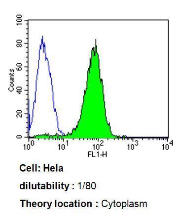
- Flow cytometry analysis of ADP Ribosylation Factor in HeLa cells compared to an isotype control (blue). Cells were harvested, adjusted to a concentration of 1-5x10^6 cells/ml, fixed with 2% paraformaldehyde, washed with PBS, and incubated with ADP Ribosylation Factor antibody [1D9] at a dilution of 1:80 for 60 min at room temperature. Cells were then blocked in a solution of 2% BSA-PBS for 30 min at room temperature, incubated for 40 min at room temperature in the dark using a proper secondary antibody, and re-suspended in PBS for FACS analysis.