Explore
 Validate
 Learn
LearnPB9336
antibody from Boster Biological Technology
Targeting: HSD17B10
17b-HSD10, ABAD, CAMR, ERAB, HADH2, MHBD, MRPP2, MRXS10, SDR5C1
 Western blot
 Immunocytochemistry
Antibody data
- Antibody Data
- Antigen structure
- References [1]
- Comments [0]
- Validations
- Western blot [1]
Submit
Validation data
Reference
Comment
Report error
- Product number
- PB9336 - Provider product page

- Provider
- Boster Biological Technology
- Product name
- Anti-ERAB/HSD17B10 Antibody Picoband™
- Antibody type
- Polyclonal
- Description
- Polyclonal antibody for ERAB/HSD17B10 detection. Host: Rabbit.Size: 100μg/vial. Tested applications: WB, IHC-P, ICC/IF, FCM. Reactive species: Human. ERAB/HSD17B10 information: Molecular Weight: 26923 MW; Subcellular Localization: Mitochondrion ; Tissue Specificity: Ubiquitously expressed in normal tissues but is overexpressed in neurons affected in AD.
- Reactivity
- Human, Mouse
- Host
- Rabbit
- Vial size
- 100μg/vial
- Concentration
- Add 0.2ml of distilled water will yield a concentration of 500ug/ml.
- Storage
- At -20°C for one year. After reconstitution, at 4°C for one month. It can also be aliquoted and stored frozen at -20°C for a longer time. Avoid repeated freezing and thawing.
- Handling
- Add 0.2ml of distilled water will yield a concentration of 500ug/ml.
Submitted references Targeting oxidative phosphorylation to increase the efficacy of immune-combination therapy in renal cell carcinoma.
Tian J, Luo J, Zeng X, Ke C, Wang Y, Liu Z, Li L, Zhang Y, Hu Z, Yang C
Journal for immunotherapy of cancer 2024 Feb 14;12(2)
Journal for immunotherapy of cancer 2024 Feb 14;12(2)
No comments: Submit comment
Supportive validation
- Submitted by
- Boster Biological Technology (provider)
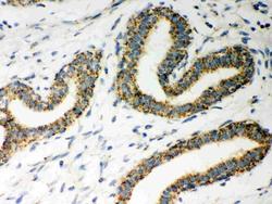
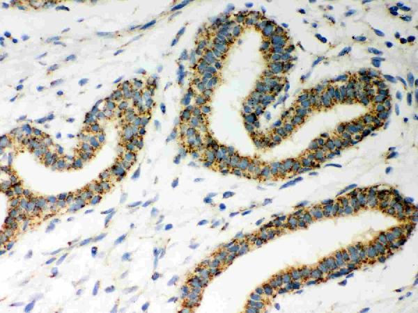
- Main image
- Experimental details
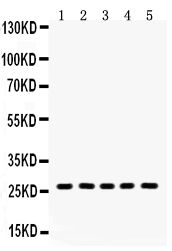
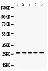
- Western blot analysis of ERAB using anti-ERAB antibody (PB9336). Electrophoresis was performed on a 5-20% SDS-PAGE gel at 70V (Stacking gel) / 90V (Resolving gel) for 2-3 hours. The sample well of each lane was loaded with 50ug of sample under reducing conditions. Lane 1: Mouse Lung Tissue Lysate, Lane 2: U87 Whole Cell Lysate, Lane 3: A549 Whole Cell Lysate, Lane 4: SW620 Whole Cell Lysate, Lane 5: 293T Whole Cell Lysate. After Electrophoresis, proteins were transferred to a Nitrocellulose membrane at 150mA for 50-90 minutes. Blocked the membrane with 5% Non-fat Milk/ TBS for 1.5 hour at RT. The membrane was incubated with rabbit anti-ERAB antigen affinity purified polyclonal antibody (Catalog # PB9336) at 0.5 μg/mL overnight at 4°C, then washed with TBS-0.1%Tween 3 times with 5 minutes each and probed with a goat anti-rabbit IgG-HRP secondary antibody at a dilution of 1:10000 for 1.5 hour at RT. The signal is developed using an Enhanced Chemiluminescent detection (ECL) kit (Catalog # EK1002) with Tanon 5200 system. A specific band was detected for ERAB at approximately 27KD. The expected band size for ERAB is at 27KD.
- Additional image