Explore
 Validate
 Learn
Learn Western blot
 Immunoprecipitation
Antibody data
- Antibody Data
- Antigen structure
- References [0]
- Comments [0]
- Validations
- Western blot [3]
Submit
Validation data
Reference
Comment
Report error
- Product number
- MA5-14852 - Provider product page

- Provider
- Invitrogen Antibodies
- Product name
- RelB Monoclonal Antibody (F.576.6)
- Antibody type
- Monoclonal
- Antigen
- Synthetic peptide
- Description
- It is not recommended to aliquot this antibody.
- Reactivity
- Human, Mouse, Rat
- Host
- Rabbit
- Isotype
- IgG
- Antibody clone number
- F.576.6
- Vial size
- 100 µL
- Concentration
- 128 µg/mL
- Storage
- -20°C
No comments: Submit comment
Supportive validation
- Submitted by
- Invitrogen Antibodies (provider)
- Main image
- Experimental details
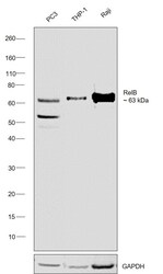
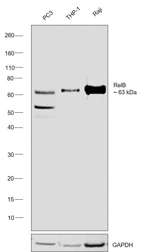
- Western blot was performed using Anti-RelB Monoclonal Antibody (F.576.6) (Product # MA5-14852) and a 63 kDa band corresponding to Transcription factor RelB was observed across across the cell lines tested. Whole cell extracts (30 µg lysate) of PC3 (Lane 1), THP-1 (Lane 2), Raji (Lane 3) were electrophoresed using NuPAGE™ 4-12% Bis-Tris Protein Gel (Product # NP0321BOX). Resolved proteins were then transferred onto a nitrocellulose membrane (Product # IB23001) by iBlot® 2 Dry Blotting System (Product # IB21001). The blot was probed with the primary antibody (1:1000 dilution) and detected by chemiluminescence with Goat anti-Rabbit IgG (H+L) Superclonal™ Recombinant Secondary Antibody, HRP (Product # A27036,1:10,000 dilution using the iBright™ FL1500 Imaging System (Product # A44115). Chemiluminescentdetection was performed using Novex® ECL Chemiluminescent Substrate Reagent Kit (Product # WP20005).
- Submitted by
- Invitrogen Antibodies (provider)
- Main image
- Experimental details
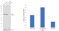
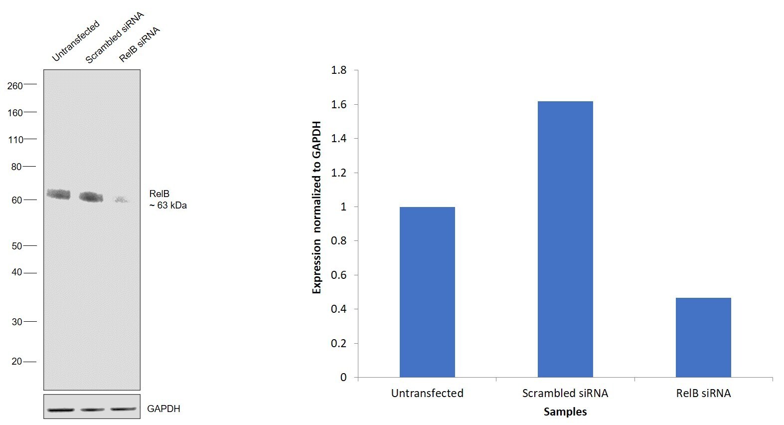
- Knockdown of Transcription factor RelB was achieved by transfecting PC-3 with Transcription factor RelB specific siRNAs (Silencer® select Product # S11917, S11919). Western blot analysis (Fig. a) was performed using Whole cell extracts from the Transcription factor RelB knockdown cells (lane 3), non-targeting scrambled siRNA transfected cells (lane 2) and untransfected cells (lane 1). The blot was probed with RelB Monoclonal Antibody (F.576.6) (Product # MA5-14852, 1:1000 dilution ) and Goat anti-Rabbit IgG (H+L) Superclonal™ Recombinant Secondary Antibody, HRP (Product # A27036, 1:10000 dilution). Densitometric analysis of this western blot is shown in histogram (Fig. b). Decrease in signal upon siRNA mediated knock down confirms that antibody is specific to Transcription factor RelB.
- Submitted by
- Invitrogen Antibodies (provider)
- Main image
- Experimental details
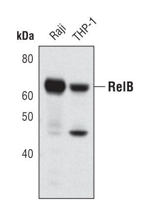
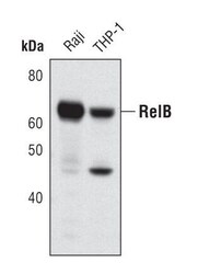
- Western blot analysis of RelB in extracts from Raji and THP-1 cells using RelB monoclonal antibody (Product # MA5-14852).