Explore
 Validate
 Learn
Learn Western blot
 Immunoprecipitation
Antibody data
- Antibody Data
- Antigen structure
- References [1]
- Comments [0]
- Validations
- Immunoprecipitation [1]
- Immunohistochemistry [2]
- Chromatin Immunoprecipitation [2]
- Other assay [1]
Submit
Validation data
Reference
Comment
Report error
- Product number
- PA5-27679 - Provider product page

- Provider
- Invitrogen Antibodies
- Product name
- RelB Polyclonal Antibody
- Antibody type
- Polyclonal
- Antigen
- Recombinant full-length protein
- Description
- Recommended positive controls: Raji, NCI-H929, NIH-3T3, RelB-transfected 293T. Predicted reactivity: Mouse (83%). Store product as a concentrated solution. Centrifuge briefly prior to opening the vial.
- Reactivity
- Human, Mouse
- Host
- Rabbit
- Isotype
- IgG
- Vial size
- 100 μL
- Concentration
- 0.42 mg/mL
- Storage
- Store at 4°C short term. For long term storage, store at -20°C, avoiding freeze/thaw cycles.
Submitted references Two-layer regulation of TRAF6 mediated by both TLR4/NF-kB signaling and miR-589-5p increases proinflammatory cytokines in the pathology of severe acute pancreatitis.
Chen Z, Dong WH, Wu Q, Wang J
American journal of translational research 2020;12(6):2379-2395
American journal of translational research 2020;12(6):2379-2395
No comments: Submit comment
Supportive validation
- Submitted by
- Invitrogen Antibodies (provider)
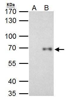
- Main image
- Experimental details
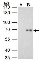
- RelB Polyclonal Antibody immunoprecipitates RelB protein in IP experiments. IP Sample: HeLa whole cell lysate/extract A. Control with 2 µg of preimmune rabbit IgG B. Immunoprecipitation of RelB protein by 2 µg of RelB Polyclonal Antibody (Product # PA5-27679) 7.5% SDS-PAGE The immunoprecipitated RelB protein was detected by RelB Polyclonal Antibody (Product # PA5-27679) diluted at 1:1,000.

Supportive validation
- Submitted by
- Invitrogen Antibodies (provider)
- Main image
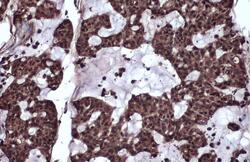
- Experimental details
- Immunohistochemistry (Paraffin) analysis of RelB was performed in paraffin-embedded human breast carcinoma tissue using RelB Polyclonal Antibody (Product # PA5-27679) at a dilution of 1:500. Antigen Retrieval: Citrate buffer, pH 6.0, 15 min.

- Submitted by
- Invitrogen Antibodies (provider)
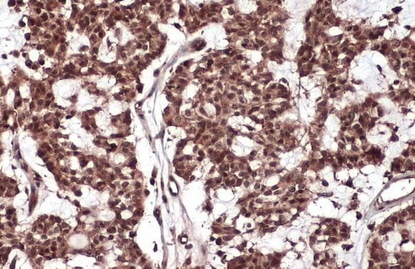
- Main image

- Experimental details
- RelB Polyclonal Antibody detects RelB protein at cytoplasm and nucleus by immunohistochemical analysis. Sample: Paraffin-embedded human breast carcinoma. RelB stained by RelB Polyclonal Antibody (Product # PA5-27679) diluted at 1:500. Antigen Retrieval: Citrate buffer, pH 6.0, 15 min.
Supportive validation
- Submitted by
- Invitrogen Antibodies (provider)
- Main image

- Experimental details
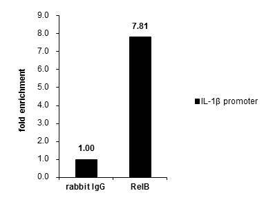
- Cross-linked ChIP was performed with THP-1 chromatin extract treated with LPS (1.0 µg/mL for 3 h) and 5 µg of either normal rabbit IgG or RelB Polyclonal Antibody (Product # PA5-27679). The precipitated DNA was detected by PCR with primer set targeting to IL-1betapromoter.

- Submitted by
- Invitrogen Antibodies (provider)
- Main image

- Experimental details
- Cross-linked ChIP was performed with THP-1 chromatin extract treated with LPS (1.0 µg/mL for 3 h) and 5 µg of either normal rabbit IgG or RelB Polyclonal Antibody (Product # PA5-27679). The precipitated DNA was detected by PCR with primer set targeting to IL-1betapromoter.
Supportive validation
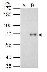
- Submitted by
- Invitrogen Antibodies (provider)
- Main image
- Experimental details
- RelB Polyclonal Antibody immunoprecipitates RelB protein in IP experiments. IP Sample: HeLa whole cell lysate/extract A. Control with 2 µg of preimmune rabbit IgG B. Immunoprecipitation of RelB protein by 2 µg of RelB Polyclonal Antibody (Product # PA5-27679) 7.5% SDS-PAGE The immunoprecipitated RelB protein was detected by RelB Polyclonal Antibody (Product # PA5-27679) diluted at 1:1,000.