Explore
 Validate
 Learn
Learn Western blot
Antibody data
- Antibody Data
- Antigen structure
- References [1]
- Comments [0]
- Validations
- Western blot [1]
- Immunocytochemistry [1]
- Immunohistochemistry [1]
Submit
Validation data
Reference
Comment
Report error
- Product number
- MAB2698 - Provider product page

- Provider
- R&D Systems
- Product name
- Human RelB Antibody
- Antibody type
- Monoclonal
- Description
- Protein A or G purified from hybridoma culture supernatant. Detects human RelB.
- Reactivity
- Human
- Host
- Mouse
- Conjugate
- Unconjugated
- Antigen sequence
Q01201- Isotype
- IgG
- Antibody clone number
- 315206
- Vial size
- 100 ug
- Concentration
- LYOPH
- Storage
- Use a manual defrost freezer and avoid repeated freeze-thaw cycles. 12 months from date of receipt, -20 to -70 °C as supplied. 1 month, 2 to 8 °C under sterile conditions after reconstitution. 6 months, -20 to -70 °C under sterile conditions after reconstitution.
Submitted references Kinome-wide functional genomics screen reveals a novel mechanism of TNFα-induced nuclear accumulation of the HIF-1α transcription factor in cancer cells.
Schoolmeesters A, Brown DD, Fedorov Y
PloS one 2012;7(2):e31270
PloS one 2012;7(2):e31270
No comments: Submit comment
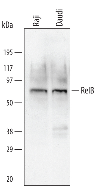
Supportive validation
- Submitted by
- R&D Systems (provider)
- Main image

- Experimental details
- Detection of Human RelB by Western Blot. Western blot shows lysates of Daudi human Burkitt's lymphoma cell line and Raji human Burkitt's lymphoma cell line. PVDF membrane was probed with 0.1 µg/mL of Mouse Anti-Human RelB Monoclonal Antibody (Catalog # MAB2698) followed by HRP-conjugated Anti-Mouse IgG Secondary Antibody (Catalog # HAF007). A specific band was detected for RelB at approximately 70 kDa (as indicated). This experiment was conducted under reducing conditions and using Immunoblot Buffer Group 4.
Supportive validation
- Submitted by
- R&D Systems (provider)
- Main image
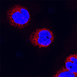
- Experimental details
- RelB in Raji Human Cell Line. RelB was detected in immersion fixed Raji human Burkitt's lymphoma cell line using Mouse Anti-Human RelB Monoclonal Antibody (Catalog # MAB2698) at 25 µg/mL for 3 hours at room temperature. Cells were stained using the NorthernLights™ 557-conjugated Anti-Mouse IgG Secondary Antibody (red; Catalog # NL007) and counterstained with DAPI (blue). Specific staining was localized to cytoplasm. View our protocol for Fluorescent ICC Staining of Non-adherent Cells.

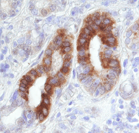
Supportive validation
- Submitted by
- R&D Systems (provider)
- Main image

- Experimental details
- RelB in Human Lymphoma. RelB was detected in immersion fixed paraffin-embedded sections of human lymphoma using 8 µg/mL Mouse Anti-Human RelB Monoclonal Antibody (Catalog # MAB2698) overnight at 4 °C. Tissue was stained with the Anti-Mouse HRP-DAB Cell & Tissue Staining Kit (brown; Catalog # CTS002) and counterstained with hematoxylin (blue). Specific labeling was localized to the cytoplasm in epithelial cells. View our protocol for Chromogenic IHC Staining of Paraffin-embedded Tissue Sections.