Explore
 Validate
 Learn
Learn Western blot
Antibody data
- Antibody Data
- Antigen structure
- References [20]
- Comments [0]
- Validations
- Western blot [2]
- Other assay [17]
Submit
Validation data
Reference
Comment
Report error
- Product number
- 44-940G - Provider product page

- Provider
- Invitrogen Antibodies
- Product name
- Phospho-PAK1/2/3 (Ser141) Polyclonal Antibody
- Antibody type
- Polyclonal
- Antigen
- Synthetic peptide
- Reactivity
- Human
- Host
- Rabbit
- Isotype
- IgG
- Vial size
- 100 µL
- Storage
- -20°C
Submitted references Phosphoproteomic of the acetylcholine pathway enables discovery of the PKC-β-PIX-Rac1-PAK cascade as a stimulatory signal for aversive learning.
Nck1, But Not Nck2, Mediates Disturbed Flow-Induced p21-Activated Kinase Activation and Endothelial Permeability.
Conditional Deletion of CC2D1A Reduces Hippocampal Synaptic Plasticity and Impairs Cognitive Function through Rac1 Hyperactivation.
Group I Paks are essential for epithelial- mesenchymal transition in an Apc-driven model of colorectal cancer.
Transgenic autoinhibition of p21-activated kinase exacerbates synaptic impairments and fronto-dependent behavioral deficits in an animal model of Alzheimer's disease.
A bead-based western for high-throughput cellular signal transduction analyses.
The calcium sensor Copine-6 regulates spine structural plasticity and learning and memory.
The matrikine N-α-PGP couples extracellular matrix fragmentation to endothelial permeability.
Group I Paks as therapeutic targets in NF2-deficient meningioma.
Pharmacological stimulation of the brain serotonin receptor 7 as a novel therapeutic approach for Rett syndrome.
Lis1 mediates planar polarity of auditory hair cells through regulation of microtubule organization.
Glucocorticoid receptors are localized to dendritic spines and influence local actin signaling.
Physiological activation of synaptic Rac>PAK (p-21 activated kinase) signaling is defective in a mouse model of fragile X syndrome.
Cytoskeletal changes underlie estrogen's acute effects on synaptic transmission and plasticity.
Exercise improves cognition and hippocampal plasticity in APOE epsilon4 mice.
Loss of modifier of cell adhesion reveals a pathway leading to axonal degeneration.
Signal transduction in Alzheimer disease: p21-activated kinase signaling requires C-terminal cleavage of APP at Asp664.
Changes in synaptic morphology accompany actin signaling during LTP.
Brain-derived neurotrophic factor promotes long-term potentiation-related cytoskeletal changes in adult hippocampus.
p21-activated kinase regulates endothelial permeability through modulation of contractility.
Yamahashi Y, Lin YH, Mouri A, Iwanaga S, Kawashima K, Tokumoto Y, Watanabe Y, Faruk MO, Zhang X, Tsuboi D, Nakano T, Saito N, Nagai T, Yamada K, Kaibuchi K
Molecular psychiatry 2022 Aug;27(8):3479-3492
Molecular psychiatry 2022 Aug;27(8):3479-3492
Nck1, But Not Nck2, Mediates Disturbed Flow-Induced p21-Activated Kinase Activation and Endothelial Permeability.
Alfaidi M, Bhattarai U, Orr AW
Journal of the American Heart Association 2020 Jun 2;9(11):e016099
Journal of the American Heart Association 2020 Jun 2;9(11):e016099
Conditional Deletion of CC2D1A Reduces Hippocampal Synaptic Plasticity and Impairs Cognitive Function through Rac1 Hyperactivation.
Yang CY, Yu TH, Wen WL, Ling P, Hsu KS
The Journal of neuroscience : the official journal of the Society for Neuroscience 2019 Jun 19;39(25):4959-4975
The Journal of neuroscience : the official journal of the Society for Neuroscience 2019 Jun 19;39(25):4959-4975
Group I Paks are essential for epithelial- mesenchymal transition in an Apc-driven model of colorectal cancer.
Chow HY, Dong B, Valencia CA, Zeng CT, Koch JN, Prudnikova TY, Chernoff J
Nature communications 2018 Aug 27;9(1):3473
Nature communications 2018 Aug 27;9(1):3473
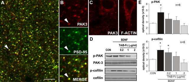
Transgenic autoinhibition of p21-activated kinase exacerbates synaptic impairments and fronto-dependent behavioral deficits in an animal model of Alzheimer's disease.
Bories C, Arsenault D, Lemire M, Tremblay C, De Koninck Y, Calon F
Aging 2017 May 16;9(5):1386-1403
Aging 2017 May 16;9(5):1386-1403
A bead-based western for high-throughput cellular signal transduction analyses.
Treindl F, Ruprecht B, Beiter Y, Schultz S, Döttinger A, Staebler A, Joos TO, Kling S, Poetz O, Fehm T, Neubauer H, Kuster B, Templin MF
Nature communications 2016 Sep 23;7:12852
Nature communications 2016 Sep 23;7:12852
The calcium sensor Copine-6 regulates spine structural plasticity and learning and memory.
Reinhard JR, Kriz A, Galic M, Angliker N, Rajalu M, Vogt KE, Ruegg MA
Nature communications 2016 May 19;7:11613
Nature communications 2016 May 19;7:11613
The matrikine N-α-PGP couples extracellular matrix fragmentation to endothelial permeability.
Hahn CS, Scott DW, Xu X, Roda MA, Payne GA, Wells JM, Viera L, Winstead CJ, Bratcher P, Sparidans RW, Redegeld FA, Jackson PL, Folkerts G, Blalock JE, Patel RP, Gaggar A
Science advances 2015;1(3)
Science advances 2015;1(3)
Group I Paks as therapeutic targets in NF2-deficient meningioma.
Chow HY, Dong B, Duron SG, Campbell DA, Ong CC, Hoeflich KP, Chang LS, Welling DB, Yang ZJ, Chernoff J
Oncotarget 2015 Feb 10;6(4):1981-94
Oncotarget 2015 Feb 10;6(4):1981-94
Pharmacological stimulation of the brain serotonin receptor 7 as a novel therapeutic approach for Rett syndrome.
De Filippis B, Nativio P, Fabbri A, Ricceri L, Adriani W, Lacivita E, Leopoldo M, Passarelli F, Fuso A, Laviola G
Neuropsychopharmacology : official publication of the American College of Neuropsychopharmacology 2014 Oct;39(11):2506-18
Neuropsychopharmacology : official publication of the American College of Neuropsychopharmacology 2014 Oct;39(11):2506-18
Lis1 mediates planar polarity of auditory hair cells through regulation of microtubule organization.
Sipe CW, Liu L, Lee J, Grimsley-Myers C, Lu X
Development (Cambridge, England) 2013 Apr;140(8):1785-95
Development (Cambridge, England) 2013 Apr;140(8):1785-95
Glucocorticoid receptors are localized to dendritic spines and influence local actin signaling.
Jafari M, Seese RR, Babayan AH, Gall CM, Lauterborn JC
Molecular neurobiology 2012 Oct;46(2):304-15
Molecular neurobiology 2012 Oct;46(2):304-15
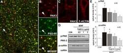
Physiological activation of synaptic Rac>PAK (p-21 activated kinase) signaling is defective in a mouse model of fragile X syndrome.
Chen LY, Rex CS, Babayan AH, Kramár EA, Lynch G, Gall CM, Lauterborn JC
The Journal of neuroscience : the official journal of the Society for Neuroscience 2010 Aug 18;30(33):10977-84
The Journal of neuroscience : the official journal of the Society for Neuroscience 2010 Aug 18;30(33):10977-84
Cytoskeletal changes underlie estrogen's acute effects on synaptic transmission and plasticity.
Kramár EA, Chen LY, Brandon NJ, Rex CS, Liu F, Gall CM, Lynch G
The Journal of neuroscience : the official journal of the Society for Neuroscience 2009 Oct 14;29(41):12982-93
The Journal of neuroscience : the official journal of the Society for Neuroscience 2009 Oct 14;29(41):12982-93
Exercise improves cognition and hippocampal plasticity in APOE epsilon4 mice.
Nichol K, Deeny SP, Seif J, Camaclang K, Cotman CW
Alzheimer's & dementia : the journal of the Alzheimer's Association 2009 Jul;5(4):287-94
Alzheimer's & dementia : the journal of the Alzheimer's Association 2009 Jul;5(4):287-94
Loss of modifier of cell adhesion reveals a pathway leading to axonal degeneration.
Chen Q, Peto CA, Shelton GD, Mizisin A, Sawchenko PE, Schubert D
The Journal of neuroscience : the official journal of the Society for Neuroscience 2009 Jan 7;29(1):118-30
The Journal of neuroscience : the official journal of the Society for Neuroscience 2009 Jan 7;29(1):118-30
Signal transduction in Alzheimer disease: p21-activated kinase signaling requires C-terminal cleavage of APP at Asp664.
Nguyen TV, Galvan V, Huang W, Banwait S, Tang H, Zhang J, Bredesen DE
Journal of neurochemistry 2008 Feb;104(4):1065-80
Journal of neurochemistry 2008 Feb;104(4):1065-80
Changes in synaptic morphology accompany actin signaling during LTP.
Chen LY, Rex CS, Casale MS, Gall CM, Lynch G
The Journal of neuroscience : the official journal of the Society for Neuroscience 2007 May 16;27(20):5363-72
The Journal of neuroscience : the official journal of the Society for Neuroscience 2007 May 16;27(20):5363-72
Brain-derived neurotrophic factor promotes long-term potentiation-related cytoskeletal changes in adult hippocampus.
Rex CS, Lin CY, Kramár EA, Chen LY, Gall CM, Lynch G
The Journal of neuroscience : the official journal of the Society for Neuroscience 2007 Mar 14;27(11):3017-29
The Journal of neuroscience : the official journal of the Society for Neuroscience 2007 Mar 14;27(11):3017-29
p21-activated kinase regulates endothelial permeability through modulation of contractility.
Stockton RA, Schaefer E, Schwartz MA
The Journal of biological chemistry 2004 Nov 5;279(45):46621-30
The Journal of biological chemistry 2004 Nov 5;279(45):46621-30
No comments: Submit comment
Supportive validation
- Submitted by
- Invitrogen Antibodies (provider)
- Main image

- Experimental details
- Western blot using PAK1/2/3 (pS141) polyclonal antibody, rabbit
- Submitted by
- Invitrogen Antibodies (provider)
- Main image

- Experimental details
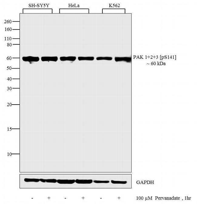
- Western blot analysis was performed on whole cell extracts (30 µg lysate) of SH-SY5Y (Lane 1), SH-SY5Y treated with 100 uM Pervanadate for 1 hour (Lane 2), HeLa (Lane 3), HeLa treated with 100 uM Pervanadate for 1 hour (Lane 4), K562 (Lane 5), K562 treated with 100 uM Pervanadate for 1 hour (Lane 6). The blots were probed with Anti-Phospho-PAK1 + 2 + 3 pSer141 Rabbit Polyclonal Antibody (Product # 44-940G, 1:500 dilution) and detected by chemiluminescence using Goat anti-Rabbit IgG (H+L) Secondary Antibody, HRP conjugate (Product # G-21234, 1:5000 dilution). A 60 kDa band corresponding to PAK1 + 2 + 3 pSer141 was observed across cell lines tested. Known quantity of protein samples were electrophoresed using Novex® NuPAGE® 12 % Bis-Tris gel (Product # NP0342BOX), XCell SureLock™ Electrophoresis System (Product # EI0002) and Novex® Sharp Pre-Stained Protein Standard (Product # LC5800). Resolved proteins were then transferred onto a nitrocellulose membrane with iBlot® 2 Dry Blotting System (Product # IB21001). The membrane was probed with the relevant primary and secondary Antibody following blocking with 5 % skimmed milk. Chemiluminescent detection was performed using Pierce™ ECL Western Blotting Substrate (Product # 32106).
Supportive validation
- Submitted by
- Invitrogen Antibodies (provider)
- Main image
- Experimental details
- NULL

- Submitted by
- Invitrogen Antibodies (provider)
- Main image

- Experimental details
- NULL
- Submitted by
- Invitrogen Antibodies (provider)
- Main image
- Experimental details
- NULL
- Submitted by
- Invitrogen Antibodies (provider)
- Main image
- Experimental details
- NULL
- Submitted by
- Invitrogen Antibodies (provider)
- Main image

- Experimental details
- NULL
- Submitted by
- Invitrogen Antibodies (provider)
- Main image
- Experimental details
- NULL
- Submitted by
- Invitrogen Antibodies (provider)
- Main image
- Experimental details
- NULL
- Submitted by
- Invitrogen Antibodies (provider)
- Main image
- Experimental details
- NULL
- Submitted by
- Invitrogen Antibodies (provider)
- Main image
- Experimental details
- NULL
- Submitted by
- Invitrogen Antibodies (provider)
- Main image
- Experimental details
- NULL
- Submitted by
- Invitrogen Antibodies (provider)
- Main image
- Experimental details
- NULL
- Submitted by
- Invitrogen Antibodies (provider)
- Main image

- Experimental details
- NULL
- Submitted by
- Invitrogen Antibodies (provider)
- Main image

- Experimental details
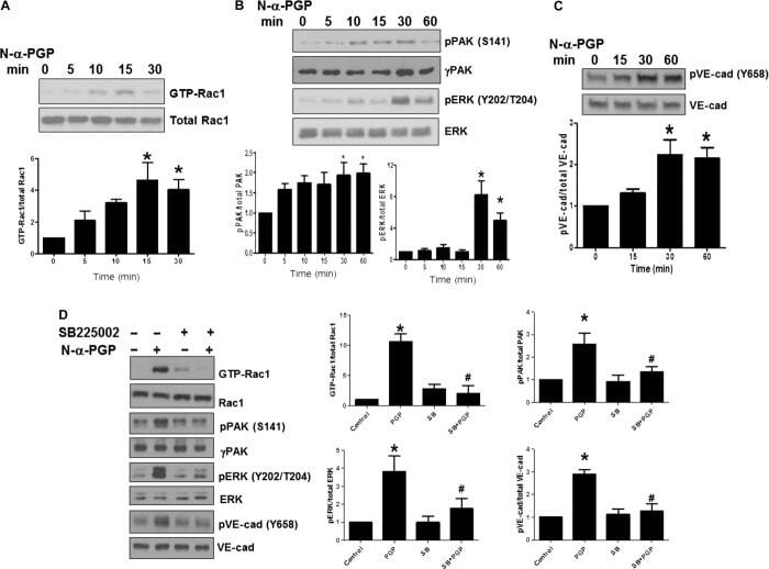
- Fig. 1 N-alpha-PGP activates endothelial cell signaling through CXCR2. ( A to C ) HUVECs were serum-starved for 2 hours before stimulation with N-alpha-PGP (0.5 mg/ml) for indicated times, and activation of Rac1 (A) and phosphorylation of PAK (pPAK) and ERK (pERK) (B) and VE-cadherin (pVE-cad) (C) were determined by Western blot. Shown are representative Western blots together with quantification. Bar graphs show means +- SEM ( n = 3). * P < 0.05 relative to time 0 by one-way analysis of variance (ANOVA) with Tukey post-test. ( D ) HUVECs were untreated or treated with N-alpha-PGP (0.5 mg/ml) (30 min) alone or after pretreatment with 200 nM SB225002, and Rac1 activity and phosphorylation of ERK, PAK, and VE-cadherin were determined by Western blot. Shown are representative Western blots together with quantification. Bar graphs show means +- SEM ( n = 3). * P < 0.05 relative to time 0, # P < 0.05 relative to PGP by one-way ANOVA with Tukey post-test.
- Submitted by
- Invitrogen Antibodies (provider)
- Main image
- Experimental details
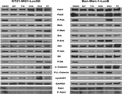
- Figure 4 The effect of Pak inhibitors on Erk and Akt-S6 signaling pathways KT21 or Ben-Men cells were treated with inhibitors for 72 hours as described in Figure 2 . Following SDS/PAGE and transfer to PVDF membranes, expression levels of Pak, Mek, Erk, Akt, S6, and beta-Catenin were assessed by immunoblot using total and phospho-specific antibodies. GADPH was used as loading control.
- Submitted by
- Invitrogen Antibodies (provider)
- Main image

- Experimental details
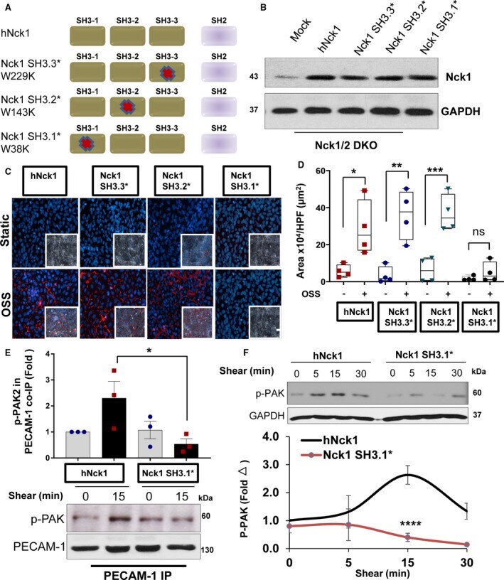
- Nck (noncatalytic region of tyrosine kinase) 1 first SH3 domain regulates PAK2 (p21-activated kinase 2)-induced endothelial permeability. A , Schematic of Nck1 point mutations in each of the SH3 domains and the nomenclature used to inactivation of SH3 domains. B , Lysates from human aortic endothelial cells (HAECs) transiently transfected with Nck1 variants with inactivated SH3 domains. C and D , Flow-induced permeability using biotinylated gelatin/Alexa 647-streptavidin assay. 4',6-Diamidino-2-phenylindole (blue; nuclei), phalloidin (green; F-actin fibers), and streptavidin (red; exposed biotinylated gelatin) Images analyzed using NIS-Elements software from n=4. Scale bar=200 mum. Data were reported as box and whiskers with scatter plots and analyzed by Kruskal-Wallis test (* P
- Submitted by
- Invitrogen Antibodies (provider)
- Main image

- Experimental details
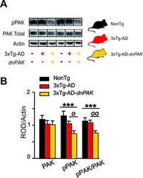
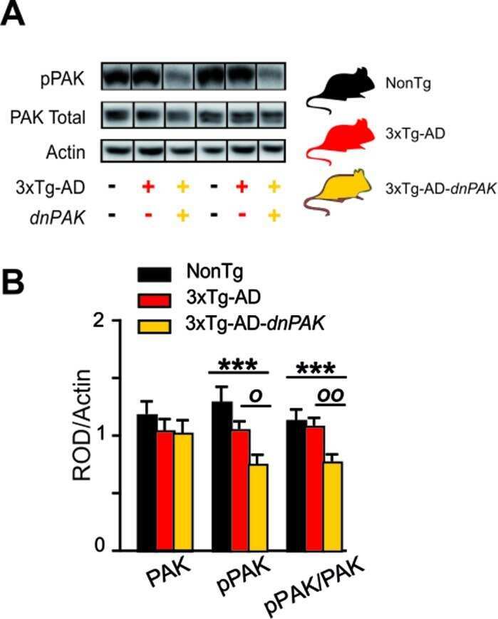
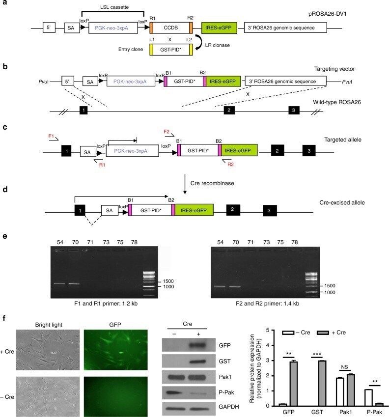
- Fig. 1 Generation and analysis of conditional Rosa26 -promoter-based expression alleles. a LR reaction performed between the pROSA26-DV1 vector and pEntry clone containing GST-PID* fragment to generate the Rosa26 targeting vector. SA is spice acceptor, PGK is phosphoglycerate kinase 1 promoter, and 3xpA is multimerized polyadenylation sequence in which loxP-PGK-neo-3xpA-loxP formed a LSL cassette. b Homologous recombination occurred between exon 1 and 2 of wild-type Rosa26 locus in G4 ES cells after electroporation. Black boxes represent the exons located at ROSA26 locus. c Knock-in targeted allele analyzed by PCR using both external primers (F1 and R1) and internal primers (F2 and R2). d Cre-mediated excision of intervening loxP flanked PGK-neo-3xpA (STOP) cassette resulted in the Rosa26 -locus-based expression of an exon1-GST-PID*-IRES-eGFP bi-cistronic fusion transcript. e Representative result of genotyping PCR analysis of tail biopsy DNA detecting the presence of fusion transcript by both external primers (F1 and R1, 1.2 kb) and internal primers (F2 and R2, 1.4 kb), in which #54 and #70 were mice revealing positive results. f Phase contrast image (left side) and green fluorescence image (right side) showing the same field of view of MEF cells derived from PID mice after Adeno-cre virus infection. Representative western blot of GFP, GST, Pak1, Phospho-Pak expression level in MEF cells after infection of Adeno-cre virus. Relative protein expression wa
- Submitted by
- Invitrogen Antibodies (provider)
- Main image
- Experimental details
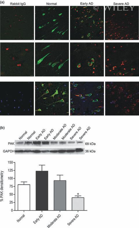
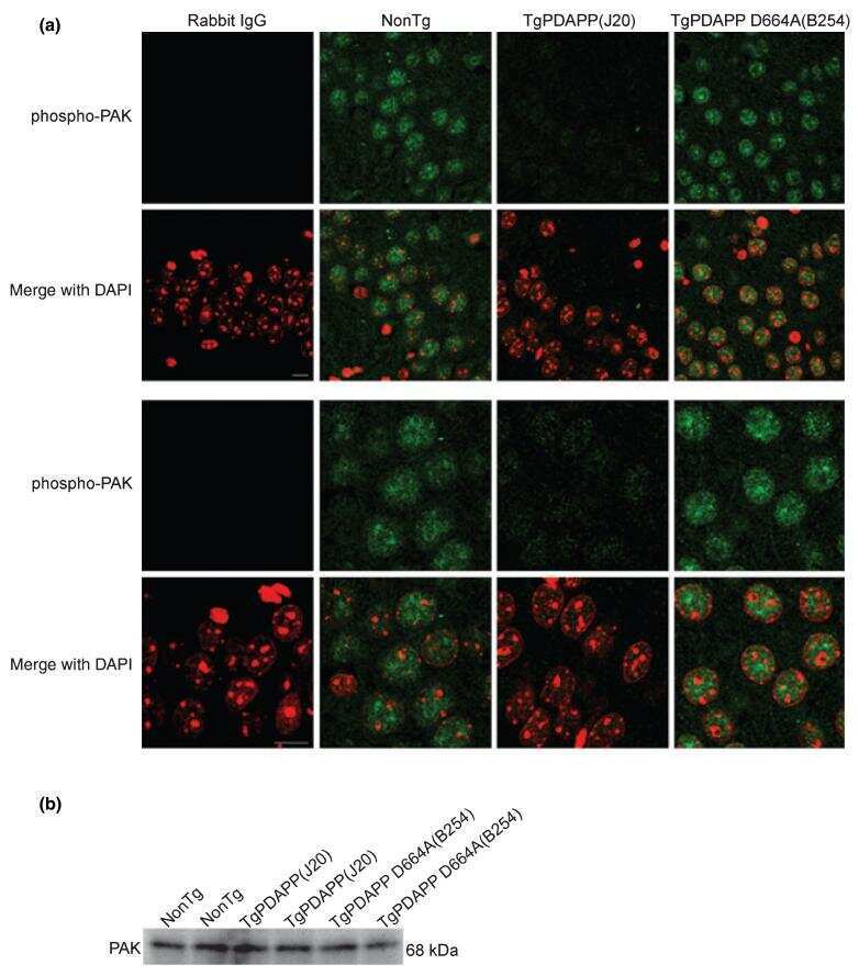
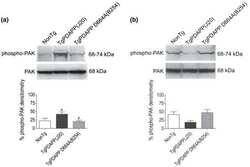
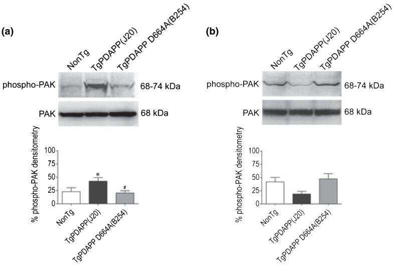
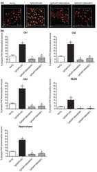
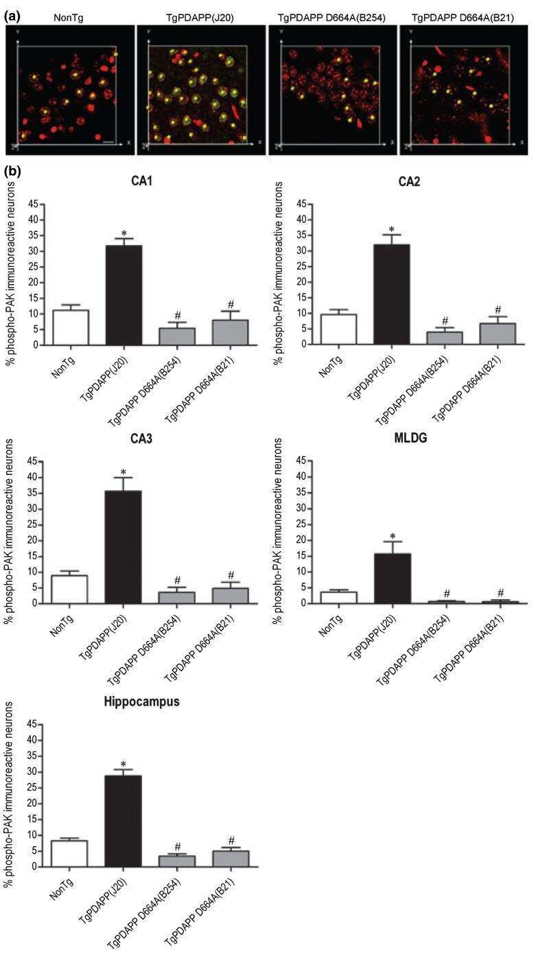
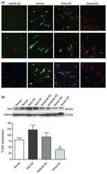
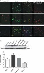
- 6 Selective changes in PAK-1/2/3 activation and expression in the hippocampus of individuals with early, moderate, and severe Alzheimer disease (AD). (a) Brain tissues from age-matched normal (control), early AD, and severe AD subjects were obtained postmortem and immunostained with an antibody against phosphorylated PAK-1/2/3 (Ser141) or rabbit IgG control, followed by Alexa Fluor488 anti-rabbit secondary (green). Nuclei were visualized with DAPI staining, pseudo-colored red. Representative images ( n = 8 human cases per group) of pyramidal hippocampal neurons were captured on a laser-scanning confocal microscope using a 40x objective and a 2x (top panel) or 4x (middle panel) zoom. Bottom panel show neurons stained with phospho-PAK-1/2/3 (green), PDI (or mouse IgG control; red), and DAPI (blue) at 40x objective and a 2x zoom. Scale bar: 10 mum. (b) A total of 100 mug each of human hippocampal extracts from normal, early AD, moderate AD, and severe AD pathology was examined by western blot using total PAK-1/2/3 (61 kDa PAK-2, 68 kDa PAK-1/3; top immunoblot) and GAPDH (36 kDa; bottom immunoblot) antibodies. Top and middle panels show representative immunoblots and the bottom panel shows mean densitometry values (+- SEM) for each group ( F = 6.3; df 3,24; p = 0.0027) of combined immunoblots ( n = 7 human cases per group). PAK-1/2/3 densitometry is expressed graphically as a percentage of the ratio of PAK to GAPDH, with denotation of significance obtained from statistical analys