 Explore
 Validate
 Learn
Learn Western blot
Antibody data
- Antibody Data
- Antigen structure
- References [0]
- Comments [0]
- Validations
- Western blot [1]
- ELISA [1]
Submit
Validation data
Reference
Comment
Report error
- Product number
- R1172 - Provider product page

- Provider
- OriGene
- Product name
- ASK1 (MAP3K5) pSer83 rabbit polyclonal antibody, Purified
- Antibody type
- Polyclonal
- Description
- ASK1 (MAP3K5) pSer83 rabbit polyclonal antibody, Purified
- Host
- Rabbit
- Conjugate
- Unconjugated
- Epitope
- MAP3K5
- Antibody clone number
- NULL
- Vial size
- 200 µg
- Concentration
- 2.0 mg/ml (by UV absorbance at 280 nm)
No comments: Submit comment
Supportive validation
- Submitted by
- OriGene (provider)
- Main image
- Experimental details
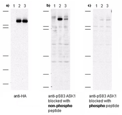
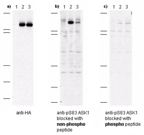
- Figure 1. Immunoblot of anti-pS83 ASK1 antibodies shows specificity for phosphorylated human ASK1. Anti-pS83 (aa 76-87) antibody, generated by immunization with phospho peptide coupled to KLH, was tested by immunoblot against lysates of Cos-7 cells after transient transfection, separately, with 1) vector only, 2) recombinant HA-ASK1, and 3) recombinant human HA-ASK1 where S83 was substituted with an alanine residue. Cells were lysed 24 h post-transfection in 200 ?L of 1x SDS-sample buffer, heated at 96°C for 5', and vortexed for 30 sec. Samples (10 ?L each) were separated on a 12% SDS-PAGE gel and transferred to PVDF (Millipore) followed by blocking for 45' using TTBS supplemented with 5% non-fat dry milk. All incubations were performed at room temperature. In panel a) all samples were incubated with 10 ug/mL mouse anti-HA antibody for 45'. After 5X washes with TTBS, reaction with ALP rabbit anti-mouse IgG at 200 ng/mL proceeded for 45' following again by washing as before. The blot was developed using BCIP/NBT. This blot demonstrates both recombinant transfections were successfully over-expressed in the Cos-7 cells. In panel b) all samples were incubated with a 1:1,000 dilution of ASK1 antibody for 45'. The antibody was pre-incubated with non-phospho peptide prior to membrane incubation. After 5X washes with TTBS, reaction with HRP goat anti-rabbit IgG at 10 ng/mL proceeded for 45' following again by washing as before. The membrane was processed as before. Lane 2 shows strong specific staining of ASK1. Lane 3, where S83 was replaced with alanine, shows greatly diminished staining. In panel c) all samples were incubated with a 1:1,000 dilution of ASK1 antibody as before except the antibody was preincubated with phospho peptide prior to membrane incubation. No staining is observed after phospho peptide blocking occurs.
- Validation comment
- WB
Supportive validation
- Submitted by
- OriGene (provider)
- Main image
- Experimental details
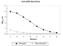
- Figure 2. ELISA results of purified polyclonal anti-pS83 ASK-1 (aa 76-87) antibody tested against BSA conjugates of non-phospho and phospho forms of immunizing peptide. Each well was coated with 0.1 mg of conjugate. The starting dilution of antibody was 1:1,000 and each point on the X-axis represents a 2-fold dilution. HRP conjugated Gt-a-Rabbit IgG H&L and TMB substrate were used for detection.
- Validation comment
- ELISA
