Explore
 Validate
 Learn
Learn Western blot
 Immunocytochemistry
Antibody data
- Antibody Data
- Antigen structure
- References [3]
- Comments [0]
- Validations
- Western blot [2]
Submit
Validation data
Reference
Comment
Report error
- Product number
- AF3398 - Provider product page

- Provider
- Novus Biologicals
- Product name
- Goat Polyclonal Catalase Antibody
- Antibody type
- Polyclonal
- Description
- Antigen Affinity-purified. Detects human, mouse and rat Catalase in Western blots.
- Reactivity
- Human, Mouse, Rat
- Host
- Goat
- Conjugate
- Unconjugated
- Isotype
- IgG
- Vial size
- 100 ug
- Concentration
- LYOPH
- Storage
- Use a manual defrost freezer and avoid repeated freeze-thaw cycles. 12 months from date of receipt, -20 to -70 degreesC as supplied. 1 month, 2 to 8 degreesC under sterile conditions after reconstitution. 6 months, -20 to -70 degreesC under sterile conditions after reconstitution.
Submitted references Mitochondrial function in liver cells is resistant to perturbations in NAD(+) salvage capacity.
A critical role of autophagy in antileukemia/lymphoma effects of APO866, an inhibitor of NAD biosynthesis.
PKG inhibits TCF signaling in colon cancer cells by blocking beta-catenin expression and activating FOXO4.
Dall M, Trammell SAJ, Asping M, Hassing AS, Agerholm M, Vienberg SG, Gillum MP, Larsen S, Treebak JT
The Journal of biological chemistry 2019 Sep 6;294(36):13304-13326
The Journal of biological chemistry 2019 Sep 6;294(36):13304-13326
A critical role of autophagy in antileukemia/lymphoma effects of APO866, an inhibitor of NAD biosynthesis.
Ginet V, Puyal J, Rummel C, Aubry D, Breton C, Cloux AJ, Majjigapu SR, Sordat B, Vogel P, Bruzzone S, Nencioni A, Duchosal MA, Nahimana A
Autophagy 2014 Apr;10(4):603-17
Autophagy 2014 Apr;10(4):603-17
PKG inhibits TCF signaling in colon cancer cells by blocking beta-catenin expression and activating FOXO4.
Kwon IK, Wang R, Thangaraju M, Shuang H, Liu K, Dashwood R, Dulin N, Ganapathy V, Browning DD
Oncogene 2010 Jun 10;29(23):3423-34
Oncogene 2010 Jun 10;29(23):3423-34
No comments: Submit comment
Supportive validation
- Submitted by
- Novus Biologicals (provider)
- Main image
- Experimental details
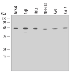
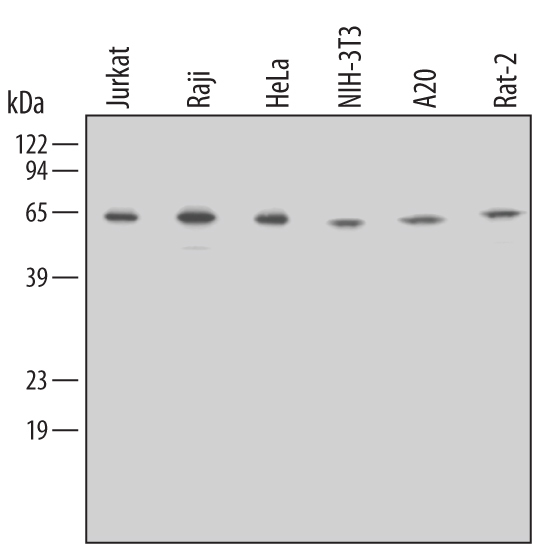
- Detection of Human/Mouse/Rat Catalase by Western Blot. Western blot shows lysates of Jurkat human acute T cell leukemia cell line, Raji human Burkitt's lymphoma cell line, HeLa human cervical epithelial carcinoma cell line, NIH-3T3 mouse embryonic fibroblast cell line, A20 mouse B cell lymphoma cell line, and Rat-2 rat embryonic fibroblast cell line. PVDF membrane was probed with 0.5 µg/mL of Goat Anti-Human/Mouse/Rat Catalase Antigen Affinity-purified Polyclonal Antibody (Catalog # AF3398) followed by HRP-conjugated Anti-Goat IgG Secondary Antibody (Catalog # HAF109). A specific band was detected for Catalase at approximately 64 kDa (as indicated). This experiment was conducted using Immunoblot Buffer Group 2.
- Submitted by
- Novus Biologicals (provider)
- Main image
- Experimental details
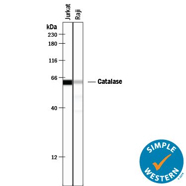
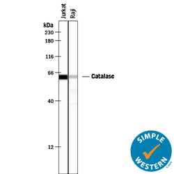
- Detection of Human Catalase by Simple WesternTM. Simple Western lane view shows lysates of Jurkat human acute T cell leukemia cell line and Raji human Burkitt's lymphoma cell line, loaded at 0.2 mg/mL. A specific band was detected for Catalase at approximately 62 kDa (as indicated) using 5 µg/mL of Goat Anti-Human/Mouse/Rat Catalase Antigen Affinity-purified Polyclonal Antibody (Catalog # AF3398) followed by 1:50 dilution of HRP-conjugated Anti-Goat IgG Secondary Antibody (Catalog # HAF109). This experiment was conducted under reducing conditions and using the 12-230 kDa separation system.