Explore
 Validate
 Learn
Learn Western blot
 Immunohistochemistry
Antibody data
- Antibody Data
- Antigen structure
- References [4]
- Comments [0]
- Validations
- Immunohistochemistry [1]
- Other assay [1]
Submit
Validation data
Reference
Comment
Report error
- Product number
- PA5-29183 - Provider product page

- Provider
- Invitrogen Antibodies
- Product name
- Catalase Polyclonal Antibody
- Antibody type
- Polyclonal
- Antigen
- Recombinant full-length protein
- Description
- Recommended positive controls: CAT-transfected 293T, mouse kidney, rat kidney. Predicted reactivity: Mouse (94%), Rat (94%), Zebrafish (88%), Drosophila (80%), Dog (97%), Pig (96%), Chicken (91%), Bovine (96%), Guinea pig (94%). Store product as a concentrated solution. Centrifuge briefly prior to opening the vial.
- Reactivity
- Human, Mouse, Rat
- Host
- Rabbit
- Isotype
- IgG
- Vial size
- 100 μL
- Concentration
- 0.47 mg/mL
- Storage
- Store at 4°C short term. For long term storage, store at -20°C, avoiding freeze/thaw cycles.
Submitted references β-Nicotinamide Mononucleotide (NMN) Administrated by Intraperitoneal Injection Mediates Protection Against UVB-Induced Skin Damage in Mice.
Ketogenic diet regulates the antioxidant catalase via the transcription factor PPARγ2.
PDGF-BB Preserves Mitochondrial Morphology, Attenuates ROS Production, and Upregulates Neuroglobin in an Astrocytic Model Under Rotenone Insult.
Nonalcoholic steatohepatitis severity is defined by a failure in compensatory antioxidant capacity in the setting of mitochondrial dysfunction.
Zhou X, Du HH, Long X, Pan Y, Hu J, Yu J, Zhao X
Journal of inflammation research 2021;14:5165-5182
Journal of inflammation research 2021;14:5165-5182
Ketogenic diet regulates the antioxidant catalase via the transcription factor PPARγ2.
Knowles S, Budney S, Deodhar M, Matthews SA, Simeone KA, Simeone TA
Epilepsy research 2018 Nov;147:71-74
Epilepsy research 2018 Nov;147:71-74
PDGF-BB Preserves Mitochondrial Morphology, Attenuates ROS Production, and Upregulates Neuroglobin in an Astrocytic Model Under Rotenone Insult.
Cabezas R, Vega-Vela NE, González-Sanmiguel J, González J, Esquinas P, Echeverria V, Barreto GE
Molecular neurobiology 2018 Apr;55(4):3085-3095
Molecular neurobiology 2018 Apr;55(4):3085-3095
Nonalcoholic steatohepatitis severity is defined by a failure in compensatory antioxidant capacity in the setting of mitochondrial dysfunction.
Boland ML, Oldham S, Boland BB, Will S, Lapointe JM, Guionaud S, Rhodes CJ, Trevaskis JL
World journal of gastroenterology 2018 Apr 28;24(16):1748-1765
World journal of gastroenterology 2018 Apr 28;24(16):1748-1765
No comments: Submit comment
Supportive validation
- Submitted by
- Invitrogen Antibodies (provider)
- Main image
- Experimental details
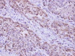
- Immunohistochemical analysis of paraffin-embedded A549 xenograft, using Catalase (Product # PA5-29183) antibody at 1:500 dilution. Antigen Retrieval: EDTA based buffer, pH 8.0, 15 min.

Supportive validation
- Submitted by
- Invitrogen Antibodies (provider)
- Main image
- Experimental details
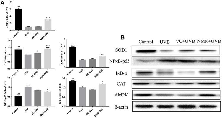
- Figure 6 Protein expression of AMPK, NFkappaB-p65, IkappaB-alpha, SOD1 and CAT in skin tissues. ( A ) relative expression levels of proteins; ( B ) protein banding map. * p < 0.05 compared to the UVB group; ** p < 0.01 compared to the UVB group; *** p < 0.001 compared to the UVB group. Abbreviations : VC+UVB, mice treated with vitamin C(300mg/kg) and UVB irradiation; NMN+UVB, mice treated with nicotinamide mononucleotide (300mg/kg) and UVB irradiation.