Explore
 Validate
 Learn
LearnMA1-23152
antibody from Invitrogen Antibodies
Targeting: ATM
ATA, ATC, ATD, ATDC, TEL1, TELO1
 Western blot
 ELISA
ELISA Immunoprecipitation Immunohistochemistry Flow cytometry Chromatin Immunoprecipitation Other assay
Antibody data
- Antibody Data
- Antigen structure
- References [3]
- Comments [0]
- Validations
- Immunoprecipitation [1]
- Immunohistochemistry [3]
- Other assay [3]
Submit
Validation data
Reference
Comment
Report error
- Product number
- MA1-23152 - Provider product page

- Provider
- Invitrogen Antibodies
- Product name
- ATM Monoclonal Antibody (2C1)
- Antibody type
- Monoclonal
- Antigen
- Recombinant full-length protein
- Description
- Recommended positive controls: SK-N-SH, HeLa, HeLa nuclear extract. Predicted molecular weight of MA1-23152 is ~350kDa. Store product as a concentrated solution. Centrifuge briefly prior to opening the vial.
- Reactivity
- Human, Mouse, Rat
- Host
- Mouse
- Isotype
- IgG
- Antibody clone number
- 2C1
- Vial size
- 100 μL
- Concentration
- 0.99 mg/mL
- Storage
- Store at 4°C short term. For long term storage, store at -20°C, avoiding freeze/thaw cycles.
Submitted references DNA-PK Inhibitor, M3814, as a New Combination Partner of Mylotarg in the Treatment of Acute Myeloid Leukemia.
Repair genes expression profile of MLH1, MSH2 and ATM in the normal oral mucosa of chronic smokers.
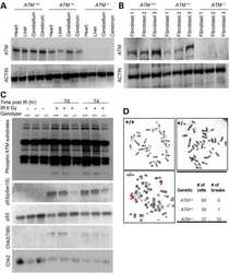
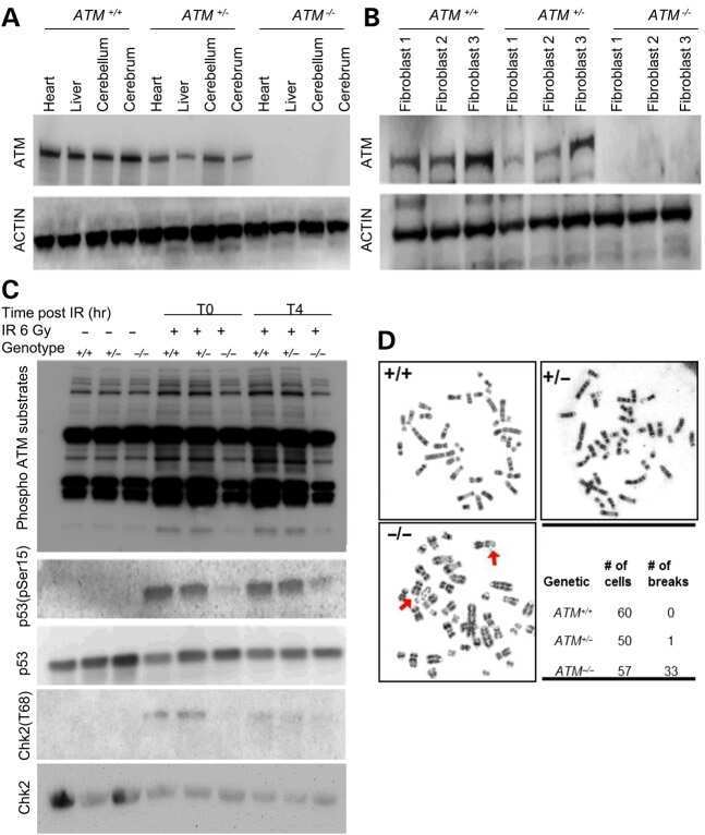
A novel porcine model of ataxia telangiectasia reproduces neurological features and motor deficits of human disease.
Carr MI, Zimmermann A, Chiu LY, Zenke FT, Blaukat A, Vassilev LT
Frontiers in oncology 2020;10:127
Frontiers in oncology 2020;10:127
Repair genes expression profile of MLH1, MSH2 and ATM in the normal oral mucosa of chronic smokers.
Alves MG, Carta CF, de Barros PP, Issa JS, Nunes FD, Almeida JD
Archives of oral biology 2017 Jan;73:60-65
Archives of oral biology 2017 Jan;73:60-65
A novel porcine model of ataxia telangiectasia reproduces neurological features and motor deficits of human disease.
Beraldi R, Chan CH, Rogers CS, Kovács AD, Meyerholz DK, Trantzas C, Lambertz AM, Darbro BW, Weber KL, White KA, Rheeden RV, Kruer MC, Dacken BA, Wang XJ, Davis BT, Rohret JA, Struzynski JT, Rohret FA, Weimer JM, Pearce DA
Human molecular genetics 2015 Nov 15;24(22):6473-84
Human molecular genetics 2015 Nov 15;24(22):6473-84
No comments: Submit comment
Supportive validation
- Submitted by
- Invitrogen Antibodies (provider)
- Main image

- Experimental details
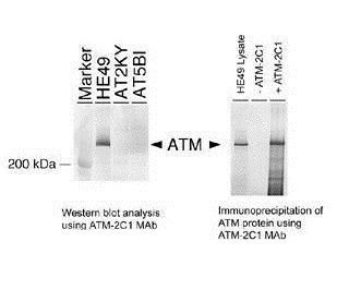
- Immunoprecipitation and Western blot of human ATM protein using anti-ATM 2C1 monoclonal antibody (Product # MA1-23152).
Supportive validation
- Submitted by
- Invitrogen Antibodies (provider)
- Main image
- Experimental details
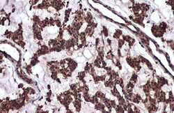
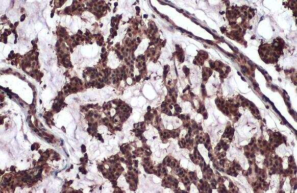
- Immunohistochemistry (Paraffin) analysis of ATM was performed in paraffin-embedded human breast carcinoma tissue using ATM Monoclonal Antibody (2C1) (Product # MA1-23152) at a dilution of 1:100. Antigen Retrieval: Citrate buffer, pH 6.0, 15 min.
- Submitted by
- Invitrogen Antibodies (provider)
- Main image
- Experimental details
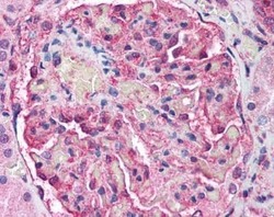
- Immunohistochemistry (Paraffin) analysis. Human Kidney (formalin-fixed, paraffin-embedded) stained with ATM Monoclonal Antibody (2C1) (Product # MA1-23152) at 5 µg/mL followed by biotinylated anti-mouse IgG secondary antibody, alkaline phosphatase-streptavidin and chromogen.

- Submitted by
- Invitrogen Antibodies (provider)
- Main image

- Experimental details
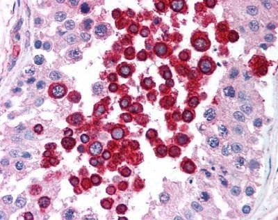
- Immunohistochemistry (Paraffin) analysis. Human Testis (formalin-fixed, paraffin-embedded) stained with ATM Monoclonal Antibody (2C1) (Product # MA1-23152) at 5 µg/mL followed by biotinylated anti-mouse IgG secondary antibody, alkaline phosphatase-streptavidin and chromogen.
Supportive validation
- Submitted by
- Invitrogen Antibodies (provider)
- Main image
- Experimental details
- NULL
- Submitted by
- Invitrogen Antibodies (provider)
- Main image
- Experimental details
- NULL
- Submitted by
- Invitrogen Antibodies (provider)
- Main image
- Experimental details
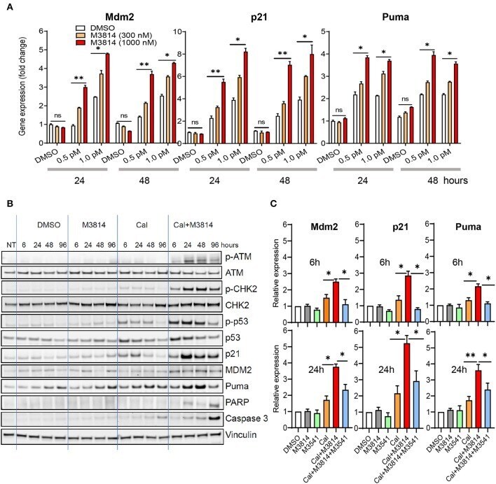
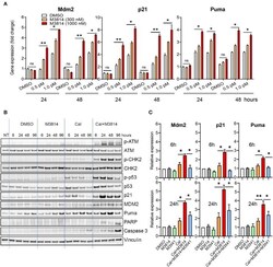
- Figure 2 M3814 overactivates p53 in response to calicheamicin in AML cells. (A) Relative gene expression analysis of key p53 transcriptional targets, Mdm2, p21 and Puma, in MV4-11 (p53 wild-type) cells treated with DMSO, calicheamicin (0.5 or 1.0 pM), or M3814 (300 or 1,000 nM) alone or in combination. Relative expression determined by the 2 (-DeltaDeltaCt) method with GAPDH reference. (B) Western blot analysis of ATM and p53 pathway proteins as well as apoptotic indicators at 6, 24, 48, and 96 h in lysates of MV4-11 cells treated with vehicle, M3814 (1 muM), calicheamicin (1pM), or the combination of calicheamicin (1 pM), and M3814 (1 muM). (C) Relative gene expression analysis at 6 and 24 h of key p53 transcriptional targets, Mdm2, p21, and Puma, in MV4-11 (p53 wild-type) cells treated with DMSO, M3814 (1 muM), M3541 (1 muM), calicheamicin (1.0 pM), calicheamicin (1 pM) + M3814 (1 muM), or calicheamicin (1 pM) + M3814 (1 muM) + M3541 (1 muM). * P < 0.05, ** P < 0.01, *** P < 0.001.